. 2019 Oct 23:10:2469.
doi: 10.3389/fimmu.2019.02469. eCollection 2019.

Pregnancy-Induced Alterations in NK Cell Phenotype and Function

Affiliations

Pregnancy-Induced Alterations in NK Cell Phenotype and Function

Mathieu Le Gars et al. Front Immunol. .

Abstract

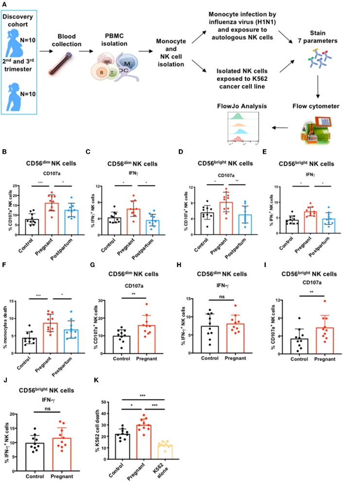
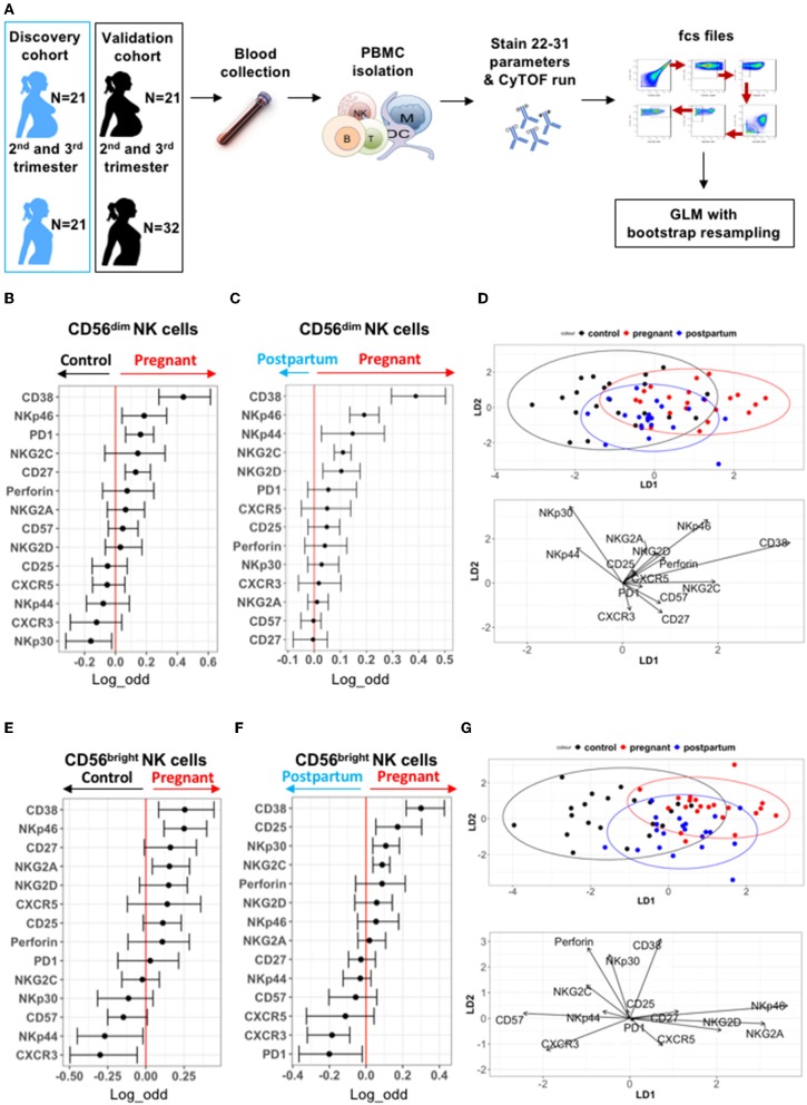
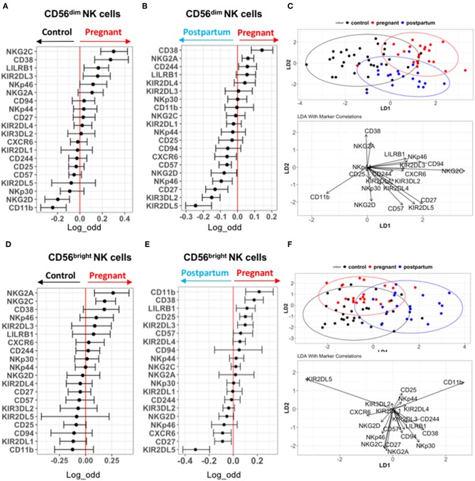
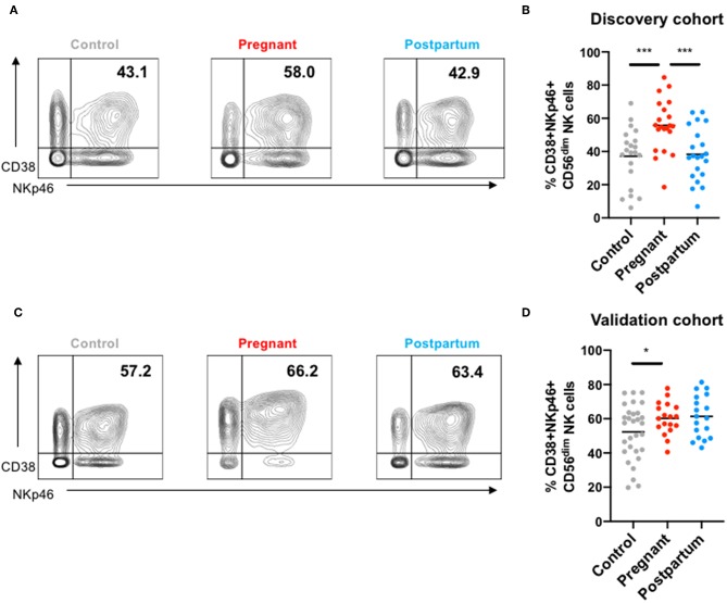
Pregnant women are particularly susceptible to complications of influenza A virus infection, which may result from pregnancy-induced changes in the function of immune cells, including natural killer (NK) cells. To better understand NK cell function during pregnancy, we assessed the ability of the two main subsets of NK cells, CD56dim, and CD56bright NK cells, to respond to influenza-virus infected cells and tumor cells. During pregnancy, CD56dim and CD56bright NK cells displayed enhanced functional responses to both infected and tumor cells, with increased expression of degranulation markers and elevated frequency of NK cells producing IFN-γ. To better understand the mechanisms driving this enhanced function, we profiled CD56dim and CD56bright NK cells from pregnant and non-pregnant women using mass cytometry. NK cells from pregnant women displayed significantly increased expression of several functional and activation markers such as CD38 on both subsets and NKp46 on CD56dim NK cells. NK cells also displayed diminished expression of the chemokine receptor CXCR3 during pregnancy. Overall, these data demonstrate that functional and phenotypic shifts occur in NK cells during pregnancy that can influence the magnitude of the immune response to both infections and tumors.

Keywords: NK cells; NK repertoire; cancer cells; influenza virus; pregnancy.


Figures

Figure 1
Figure 1
CD56dim and CD56bright NK cell immune response to influenza infected and tumor cells during pregnancy. (A) PBMCs from controls (N = 10), pregnant women (N = 10), and post-partum women (N = 10) in discovery cohort were isolated from blood samples. Monocytes and total NK cells were sorted and monocytes were infected with the H1N1 influenza virus strain. NK cells were either exposed to H1N1-infected monocytes or to K562 tumor cells for 7 or 4 h, respectively. (B–I) CD56dim and CD56bright NK cell immune response was then determined by flow cytometry. The frequency of (B) CD107a- and (C) IFN-γ-expressing CD56dim NK cells in response to influenza-infected monocytes is represented. The frequency of (D) CD107a- and (E) IFN-γ-expressing CD56bright NK cells in response to influenza-infected monocytes is represented. (F) The frequency of dead or dying monocytes based on staining with viability dye in NK cell co-culture. The frequency of CD107a (G) and IFN-γ-production (H) by CD56dim NK cells in response to K562 cells is represented. The frequency of CD107a (I) and IFN-γ-production (J) by CD56bright NK cells in response to K562 cells. (K) The frequency of dead or dying K562 tumor cells based on staining with viability dye in NK cell co-culture. *P < 0.05, **P < 0.01, and ***P < 0.001 (Mann–Whitney U-Tests to compare controls vs. pregnant; Wilcoxon matched-paired test to compare pregnant vs. post-partum).
Figure 2
Figure 2
Deep profiling of CD56dim and CD56bright NK cells in non-pregnant and pregnant women from discovery cohort. (A) PBMCs from controls (N = 21), pregnant women (N = 21), and post-partum women (N = 21) in discovery cohort were isolated from blood samples. PBMCs were isolated and stained for mass cytometry. Data obtained were analyzed using a linear regression model, GLM. (B,C) Markers predictive of CD56dim NK cells in control vs. pregnant women in discovery cohort were assessed by GLM with bootstrap resampling. The markers are listed on the y-axis and the x-axis represents the log-odds that the marker expression levels predict the outcome (control on the left vs. pregnancy on the right). Summary data are depicted, showing the 95% confidence interval. Markers in which the bar does not cross zero are significantly predictive of one state vs. the other. (D) Represents the linear discriminant analysis (LDA) for CD56dim NK cells between non-pregnant, pregnant, and post-partum samples from discovery cohort. (E,F) Markers predictive of CD56bright NK cells in control vs. pregnant women in discovery cohort were assessed by GLM with bootstrap resampling. The markers are listed on the y-axis and the x-axis represents the log-odds that the marker expression levels predict the outcome (control on the left vs. pregnancy on the right). Summary data are depicted, showing the 95% confidence interval. Markers in which the bar does not cross zero are significantly predictive of one state vs. the other. (G) Represents the linear discriminant analysis (LDA) for CD56bright NK cells between non-pregnant, pregnant, and post-partum samples from discovery cohort.
Figure 3
Figure 3
Deep profiling of CD56dim and CD56bright NK cells in non-pregnant and pregnant women from validation cohort. (A,B) Markers predictive of CD56dim NK cells in control vs. pregnant women in validation cohort were assessed by GLM with bootstrap resampling. The markers are listed on the y-axis and the x-axis represents the log-odds that the marker expression levels predict the outcome (control on the left vs. pregnancy on the right). Summary data are depicted, showing the 95% confidence interval. Markers in which the bar does not cross zero are significantly predictive of one state vs. the other. (C) Represents the linear discriminant analysis (LDA) for CD56dim NK cells between non-pregnant, pregnant and post-partum samples from validation cohort. (D,E) Markers predictive of CD56bright NK cells in control vs. pregnant women in validation cohort were assessed by GLM with bootstrap resampling. The markers are listed on the y-axis and the x-axis represents the log-odds that the marker expression levels predict the outcome (control on the left vs. pregnancy on the right). Summary data are depicted, showing the 95% confidence interval. Markers in which the bar does not cross zero are significantly predictive of one state vs. the other. (F) Represents the linear discriminant analysis (LDA) for CD56bright NK cells between non-pregnant, pregnant and post-partum samples from validation cohort.
Figure 4
Figure 4
NKp46 and CD38 co-expression in NK cells from controls, pregnant and post-partum women in the discovery and validation cohort. (A) Representative mass cytometry plots showing the co-expression of NKp46 and CD38 in peripheral NK cells from a control, pregnant or post-partum women in the discovery cohort. (B) Frequency of CD38+NKp46+ NK cells from controls, pregnant and post-partum individuals in the discovery cohort. ***P < 0.001 (Mann–Whitney U-Tests to compare controls vs. pregnant; Wilcoxon matched-paired test to compare pregnant vs. post-partum). (C) Representative mass cytometry plots showing the co-expression of NKp46 and CD38 in peripheral NK cells from a control, pregnant, or post-partum women in the validation cohort. (D) Frequency of CD38+NKp46+ NK cells from controls, pregnant and post-partum individuals in the validation cohort. *P < 0.05 (Mann–Whitney U-Tests to compare controls vs. pregnant; Wilcoxon matched-paired test to compare pregnant vs. post-partum).

References

    1. Erlebacher A. Immunology of the maternal-fetal interface. Ann Rev Immunol. (2013) 31:387–411. 10.1146/annurev-immunol-032712-100003 - DOI - PubMed
    1. Kourtis AP, Read JS, Jamieson DJ. Pregnancy and infection. N Engl J Med. (2014) 370:2211–8. 10.1056/NEJMra1213566 - DOI - PMC - PubMed
    1. Periolo N, Avaro M, Czech A, Russo M, Benedetti E, Pontoriero A, et al. . Pregnant women infected with pandemic influenza A(H1N1)pdm09 virus showed differential immune response correlated with disease severity. J Clin Virol. (2015) 64:52–8. 10.1016/j.jcv.2015.01.009 - DOI - PubMed
    1. PrabhuDas M, Bonney E, Caron K, Dey S, Erlebacher A, Fazleabas A, et al. . Immune mechanisms at the maternal-fetal interface: perspectives and challenges. Nat Immunol. (2015) 16:328–34. 10.1038/ni.3131 - DOI - PMC - PubMed
    1. Littauer EQ, Esser ES, Antao OQ, Vassilieva EV, Compans RW, Skountzou I. H1N1 influenza virus infection results in adverse pregnancy outcomes by disrupting tissue-specific hormonal regulation. PLoS Pathogens. (2017) 13:e1006757. 10.1371/journal.ppat.1006757 - DOI - PMC - PubMed

Publication types