Review
. 2019 Oct 23:10:2520.
doi: 10.3389/fimmu.2019.02520. eCollection 2019.

Development of Unconventional T Cells Controlled by MicroRNA

Affiliations
Review

Development of Unconventional T Cells Controlled by MicroRNA

Samantha J Winter et al. Front Immunol. .

Abstract

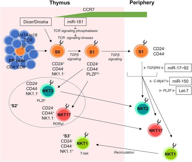
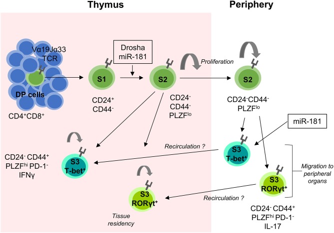
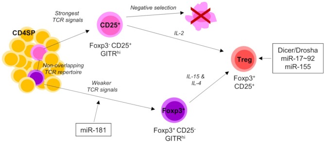
Post-transcriptional gene regulation through microRNA (miRNA) has emerged as a major control mechanism of multiple biological processes, including development and function of T cells. T cells are vital components of the immune system, with conventional T cells playing a central role in adaptive immunity and unconventional T cells having additional functions reminiscent of both innate and adaptive immunity, such as involvement in stress responses and tissue homeostasis. Unconventional T cells encompass cells expressing semi-invariant T cell receptors (TCRs), such as invariant Natural Killer T (iNKT) and Mucosal-Associated Invariant T (MAIT) cells. Additionally, some T cells with diverse TCR repertoires, including γδT cells, intraepithelial lymphocytes (IEL) and regulatory T (Treg) cells, share some functional and/or developmental features with their semi-invariant unconventional counterparts. Unconventional T cells are particularly sensitive to disruption of miRNA function, both globally and on the individual miRNA level. Here, we review the role of miRNA in the development and function of unconventional T cells from an iNKT-centric point of view. The function of single miRNAs can provide important insights into shared and individual pathways for the formation of different unconventional T cell subsets.

Keywords: MAIT cell; NKT cell; T cell; Treg cell; miR-181; microRNA; thymus; γδT cell.


Figures

Figure 1
Figure 1
miRNA-mediated control of iNKT-cell development. iNKT cells are selected on CD1d-expressing DP cells in the thymus. Subsequently they progress through distinct developmental stages S0–S3 characterized by their dynamic expression of CD24, CD44, NK1.1, and the transcription factors PLZF, RORγt, and T-bet. Early stage iNKT cells are sensitive to the loss of total miRNAs in Dicer and/or Drosha-deficient mice and require fine-tuned TGFβ and TCR signaling for normal development. S1 cells begin to upregulate the chemokine receptor CCR7, which promotes migration to the medulla and is detectable on the surface of recent thymic emigrants (RTEs). S2 cells exit the thymus and travel to peripheral tissues, whilst S3 cells recirculate back into the thymus and remain resident for extended periods of time. Individual miRNAs acting on distinct developmental transitions and their targets are indicated. Double positive (DP), Stage 0 (S0), Stage 1 (S1), Stage 2 (S2), and Stage 3 (S3).
Figure 2
Figure 2
miRNA-mediated control of MAIT-cell development. MAIT cells are selected on MR1-expressing DP thymic cells. They proceed through several stages of development (S1–S3) as indicated by their surface expression of CD24 and CD44. The transition between S1 and S2 is heavily dependent on the presence of miRNAs as shown in Drosha-deficient mouse models. S2 MAIT cells begin to proliferate and develop into S3 effector MAIT cells defined by their expression of the transcription factors T-bet and RORγt. S2 cells can also exit the thymus, where they continue to proliferate and develop into S3 MAIT cells and migrate to peripheral tissues. It is postulated that some extra-thymically matured MAIT cells recirculate back to the thymus where they contribute to the thymic microenvironment and remain resident for extended periods of time. miR-181 is currently the only miRNA shown to be involved in MAIT cell development as indicated. MHC-related protein 1 (MR1), Double positive (DP), Stage 1 (S1), Stage 2 (S2), and Stage 3 (S3).
Figure 3
Figure 3
miRNA-mediated control of Treg-cell development. Thymic-derived Treg-cell development begins at the CD4 SP stage of development and proceeds via one of two different pathways. The first Treg precursors to arise are the CD25+ population. CD25+ precursors require strong TCR signals and IL-2 signaling to mature into CD25+Foxp3+ Tregs. The alternative pathway of Treg-cell development is characterized by the generation of Foxp3+ precursors. Foxp3+ precursors develop from comparably weaker TCR signals to CD25+ precursors and share little TCR repertoire overlap. As they mature into Tregs they upregulate CD25 and require both IL-4 and IL-15 signaling. Mature Tregs emigrate from the thymus to join the peripheral T cell pool. Total (Dicer/Drosha) and individual miRNAs acting on distinct developmental transitions are indicated. Single positive (SP) and T cell receptor (TCR).

References

    1. Godfrey DI, Uldrich AP, McCluskey J, Rossjohn J, Moody DB. The burgeoning family of unconventional T cells. Nat Immunol. (2015) 16:1114–23. 10.1038/ni.3298 - DOI - PubMed
    1. Legoux F, Salou M, Lantz O. Unconventional or preset αβ T cells: evolutionarily conserved tissue-resident T cells recognizing nonpeptidic ligands. Annu Rev Cell Dev Biol. (2017) 33:511–35. 10.1146/annurev-cellbio-100616-060725 - DOI - PubMed
    1. McDonald BD, Jabri B, Bendelac A. Diverse developmental pathways of intestinal intraepithelial lymphocytes. Nat Rev Immunol. (2018) 18:514–25. 10.1038/s41577-018-0013-7 - DOI - PMC - PubMed
    1. Crosby CM, Kronenberg M. Tissue-specific functions of invariant natural killer T cells. Nat Rev Immunol. (2018) 18:559–74. 10.1038/s41577-018-0034-2 - DOI - PMC - PubMed
    1. Moran AE, Holzapfel KL, Xing Y, Cunningham NR, Maltzman JS, Punt J, et al. . T cell receptor signal strength in Treg and iNKT cell development demonstrated by a novel fluorescent reporter mouse. J Exp Med. (2011) 208:1279–89. 10.1084/jem.20110308 - DOI - PMC - PubMed

Publication types

LinkOut - more resources