

Case Reports
doi: 10.1016/j.rmcr.2019.100949.
eCollection 2019.
Two cases of primary human parechovirus pneumonia in adults
Affiliations
- PMID: 31709139
- PMCID: PMC6831859
- DOI: 10.1016/j.rmcr.2019.100949
Item in Clipboard
Case Reports
Two cases of primary human parechovirus pneumonia in adults
Respir Med Case Rep.
.
Abstract
Human parechoviruses (HPeV) are mainly isolated from upper respiratory tract infection and gastroenteritis in children. HPeV has not been screened for in the past studies of community-acquired pneumonia (CAP) in adults, and its association with CAP is unknown. We present two cases that HPeV was detected by multiplex polymerase chain reaction for respiratory viruses using bronchoalveolar lavage fluid and diagnosed as pneumonia caused by HPeV.
Keywords: Bronchoalveolar lavage fluid; HPeV; PCR; Viral pneumonia; human parechovirus.
© 2019 The Authors.
Conflict of interest statement
None.
Figures
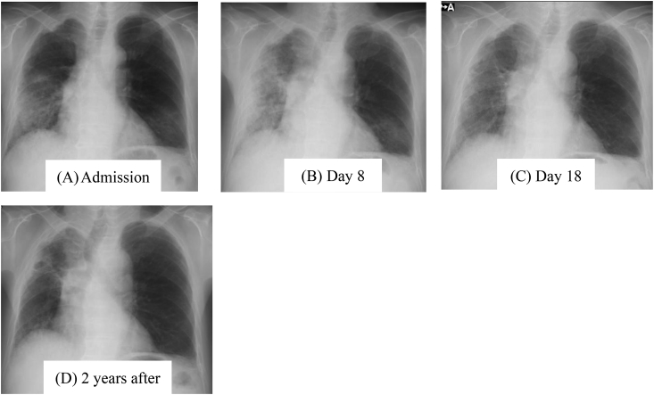
Chest X-rays in case 1. Chest X-ray on admission (A) showed consolidation and reduced volume of the right lung. The greatest deterioration had occurred on day 8 (B), and by the time of discharge on day 18, they had improved but were still somewhat present (C). The reduced volume of the right lung has remained after two years (D).
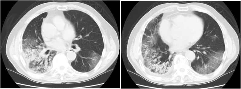
Chest computed tomography (CT) in case 1. Chest CT on admission showed bilateral consolidation (right dominant), ground-glass opacities (GGOs) around the consolidation, and air-bronchogram accompanying traction bronchiectasis within the consolidation. The GGOs in part showed non-segmental distribution.
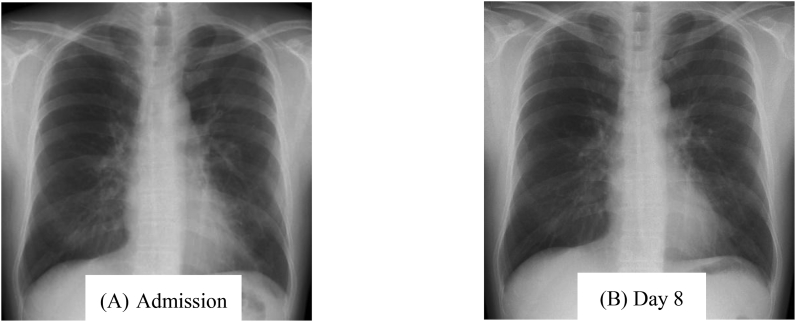
Chest X-rays in case 2. Chest X-ray on admission (A) showed nodular consolidation on both sides of the lung that had almost resolved at day 8 (B).
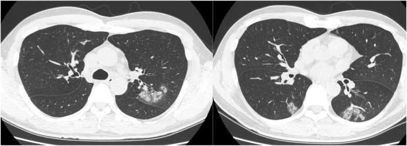
Chest computed tomography (CT) in case 2. Chest CT on admission showed patchy consolidation and GGOs along the bronchial vascular bundle in the upper and lower lobes of the left lung and upper segment of the right lower lobe. Traction bronchiectasis and volume reduction of the lungs were not observed.
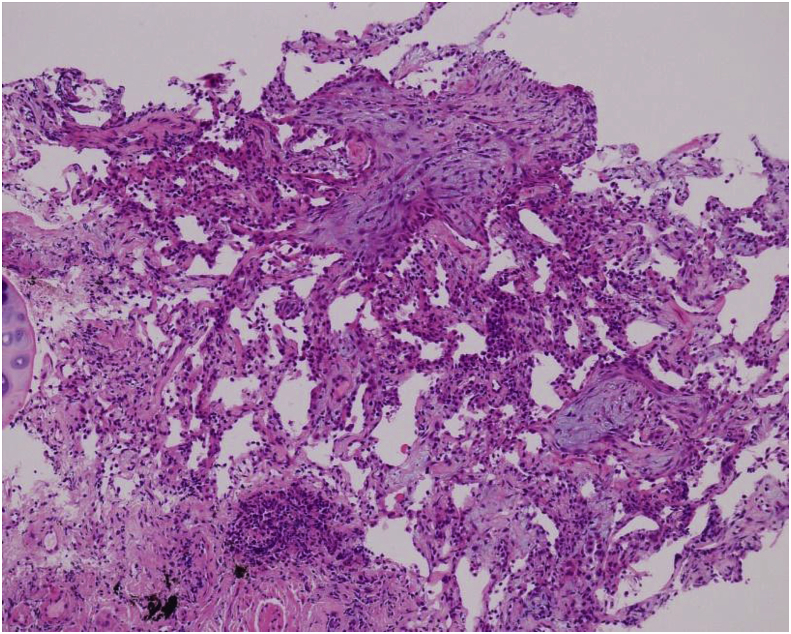
Histologic findings. Histologic findings from transbronchial lung biopsy in case 2 showed organization, swollen pneumocytes, and alveolar septal thickening with inflammatory cells (hematoxylin and eosin staining; magnification, × 50).
Similar articles
-
Diversity of human parechoviruses in Bulgaria, 2011: Detection of rare genotypes 8 and 10.Infect Genet Evol. 2015 Dec;36:315-322. doi: 10.1016/j.meegid.2015.10.004. Epub 2015 Oct 9. Infect Genet Evol. 2015. PMID: 26453770
-
Human parechovirus type 3 central nervous system infections in Israeli infants.J Clin Virol. 2013 Sep;58(1):205-10. doi: 10.1016/j.jcv.2013.06.001. Epub 2013 Jun 28. J Clin Virol. 2013. PMID: 23810613
-
Detection of human parechoviruses in children with gastroenteritis in South Korea.Arch Virol. 2011 Aug;156(8):1471-5. doi: 10.1007/s00705-011-0995-y. Epub 2011 Apr 11. Arch Virol. 2011. PMID: 21479947
-
Human parechovirus-3 infection in children, South Korea.J Clin Virol. 2013 Sep;58(1):194-9. doi: 10.1016/j.jcv.2013.05.023. Epub 2013 Jun 22. J Clin Virol. 2013. PMID: 23800693
-
Pediatric parechovirus infections.J Clin Virol. 2014 Jun;60(2):84-9. doi: 10.1016/j.jcv.2014.03.003. Epub 2014 Mar 13. J Clin Virol. 2014. PMID: 24690382 Review.
Cited by
-
Genome sequencing and phylogenetic reconstruction reveal a potential fourth rhinovirus species and its worldwide distribution.Arch Virol. 2021 Jan;166(1):225-229. doi: 10.1007/s00705-020-04855-5. Epub 2020 Oct 21. Arch Virol. 2021. PMID: 33084935
References
-
- Stanway G., Joki-Korpela P., Hyypiä T. Human parechoviruses-biology and clinical significance. Rev. Med. Virol. 2000;10(1):57–69. - PubMed
-
- Wigand R., Sabin A.B. Properties of ECHO types 22, 23 and 24. Arch. Gesamte Virusforsch. 1961;11:224–247. - PubMed
-
- Harvala H., Simmonds P. Human parechoviruses: biology, epidemiology and clinical significance. J. Clin. Virol. 2009;45:1–9. - PubMed
-
- Mizuta K., Kuroda M., Kurimura M., Yahata Y., Sekizuka T., Aoki Y., Ikeda T., Abiko C., Noda M., Kimura H., Mizutani T., Kato T., Kawanami T., Ahiko T. Epidemic myalgia in adults associated with human parechovirus type 3 infection, Yamagata, Japan. Emerg. Infect. Dis. 2008;18:1787–1793. 2012. - PMC - PubMed
Publication types
LinkOut - more resources
Full Text Sources
Miscellaneous
