

Renal actinomycosis presenting as uro-cutaneous fistula
- PMID: 31709154
- PMCID: PMC6833357
- DOI: 10.1016/j.eucr.2019.101054
Renal actinomycosis presenting as uro-cutaneous fistula
Abstract
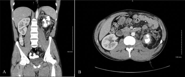
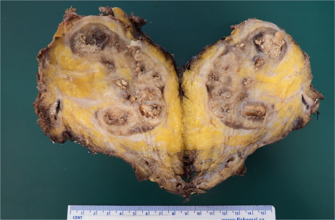
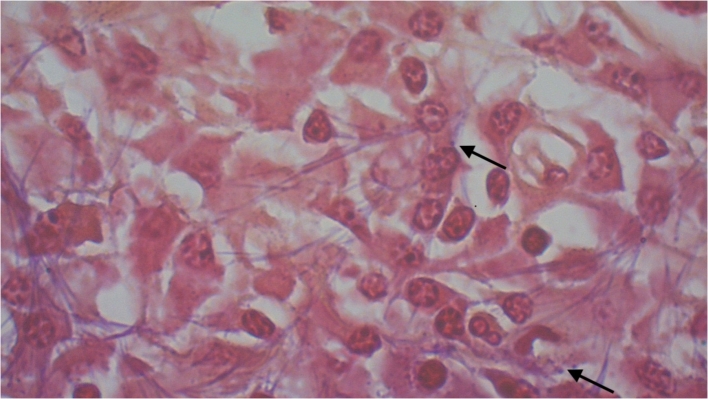
Renal actinomycosis is a rare clinical entity. Diagnosis is usually made after resection. A 36-year-old male presented with uro-cutaneous fistula and left xanthogranulomatous pyelonephritis. He was offered left open radical nephrectomy. Intra-operatively, there was "woody" inflammation of the left kidney fistulizing to the splenic flexure of the colon. We successfully resected it and a segment of the colon that had fistulized. His tissue cultures grew Actinomyces odontolyticus. Post-operatively, he received 6 weeks of intravenous beta-lactam antibiotic. He recovered well without any complications. In conclusion, renal actinomycosis can be challenging to diagnose, operate and eradicate. Perioperative considerations are presented for successful management.
Keywords: Actinomycosis; Disease management; Infection; Kidney calculi; Pyelonephritis xanthogranulomatous; Urinary fistula.
© 2019 The Authors. Published by Elsevier Inc.
Conflict of interest statement
None declared.
Figures
References
-
- Wong V.K., Turmezei T.D., Weston V.C. Actinomycosis. BMJ. 2011 Oct 11;343:d6099. - PubMed
-
- Dayan K., Neufeld D., Zissin R. Actinomycosis of the large bowel: unusual presentations and their surgical treatment. Eur J Surg. 1996;162(8):657–660. - PubMed
-
- Crosse J.E., Soderdahl D.W., Schamber D.T. Renal actinomycosis. Urology. 1976;7:309–311. - PubMed
-
- Dhanani N.N., Jones D.M., Grossman H.B. Medical management of renal actinomycosis. J Urol. 2004;171(6 Pt 1):2373–2374. - PubMed
Publication types
LinkOut - more resources
Full Text Sources
