

Case Reports
doi: 10.1016/j.case.2019.05.004.
eCollection 2019 Oct.
Forme Fruste Cor Triatriatum Dexter by Transesophageal Echocardiography and Its Impact on Percutaneous Heart Procedures: A Case Series
Affiliations
- PMID: 31709369
- PMCID: PMC6833460
- DOI: 10.1016/j.case.2019.05.004
Item in Clipboard
Case Reports
Forme Fruste Cor Triatriatum Dexter by Transesophageal Echocardiography and Its Impact on Percutaneous Heart Procedures: A Case Series
CASE (Phila).
.
No abstract available
Keywords: Congenital; Heart defects; Heart septal defects; Heart valve prosthesis implantation; Vascular access devices.
Figures

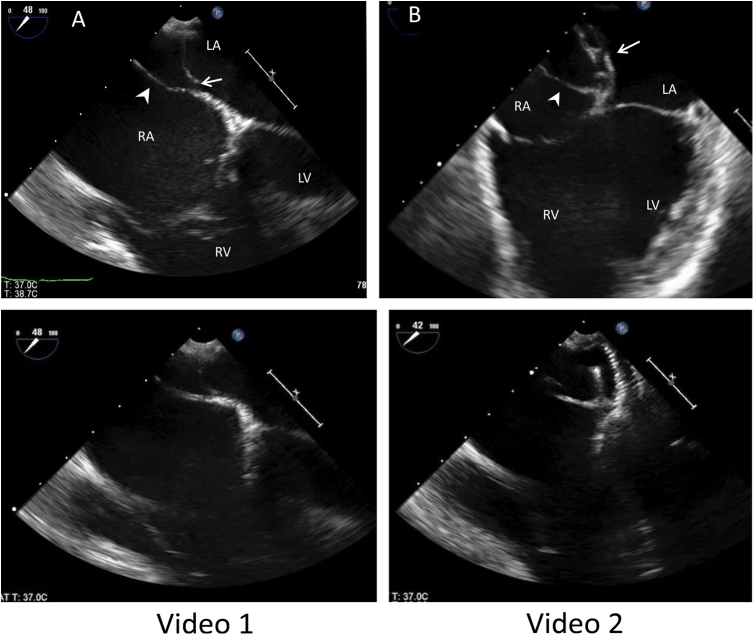
Case 1. Two-dimensional TEE images in the midesophageal four-chamber view. The ff-CTD membrane (arrowhead) is attached to the atrial septum adjacent to the fossa ovalis, producing a telltale Y sign. (A) A small secundum ASD is also shown (arrow). (B) The deployed ASD closure device (arrow). LA, Left atrium; LV, left ventricle.
Case 2. Two-dimensional TEE images in the midesophageal four-chamber view. (A) The ff-CTD membrane (arrowhead) is attached to the atrial septum adjacent to the fossa ovalis, producing a telltale Y sign. (B) PFO closure device with precarious and incomplete deployment (arrow). LA, Left atrium.
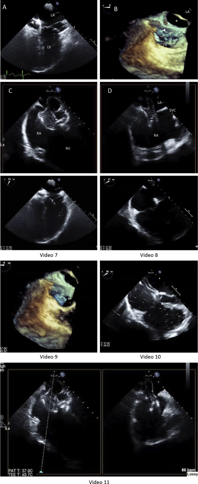
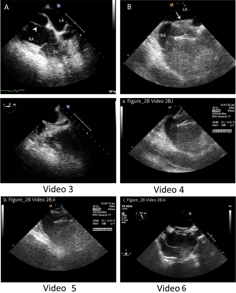
Case 3. Two-dimensional TEE images in the midesophageal four-chamber view. (A) The ff-CTD membrane (arrowheads) is attached to the atrial septum adjacent to the fossa ovalis, producing a telltale Y sign. (B) Large secundum ASD detected by 3D TEE (*). Simultaneous orthogonal plane imaging shows malpositioned Amplatzer device within a large secundum ASD in modified (C) four-chamber view and (D) bicaval view. Note the nearly 180° abnormal rotation of the closure device atrial disks, which span the ASD rather than sealing the ASD on both sides. LA, Left atrium; LV, left ventricle.
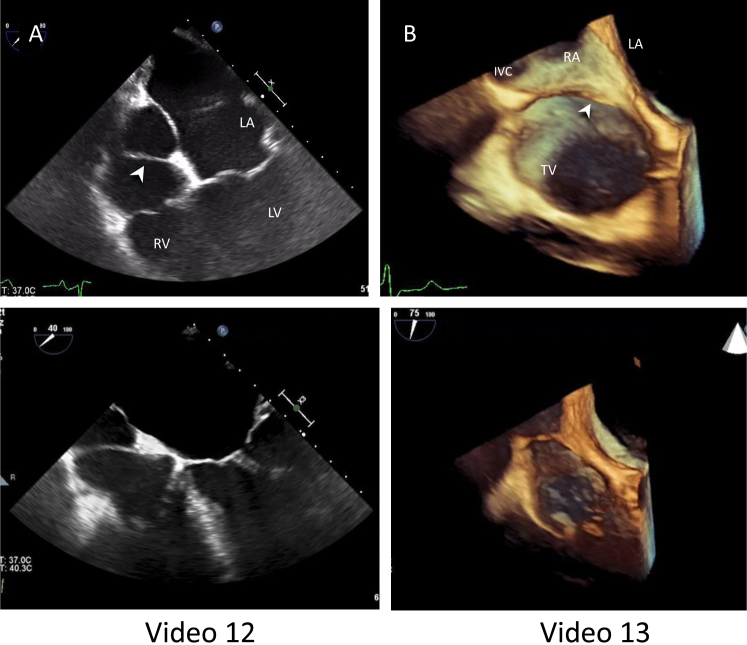
Case 4. Two-dimensional TEE image (A) in the midesophageal four-chamber view shows the ff-CTD membrane (arrowhead) attached to the atrial septum adjacent to the fossa ovalis, producing a telltale Y sign. (B) TEE 3D views of ff-CTD membrane (arrowhead) using a semi–en face view of the atrial septum and TV annulus modified to best show the defect. LA, Left atrium; LV, left ventricle.
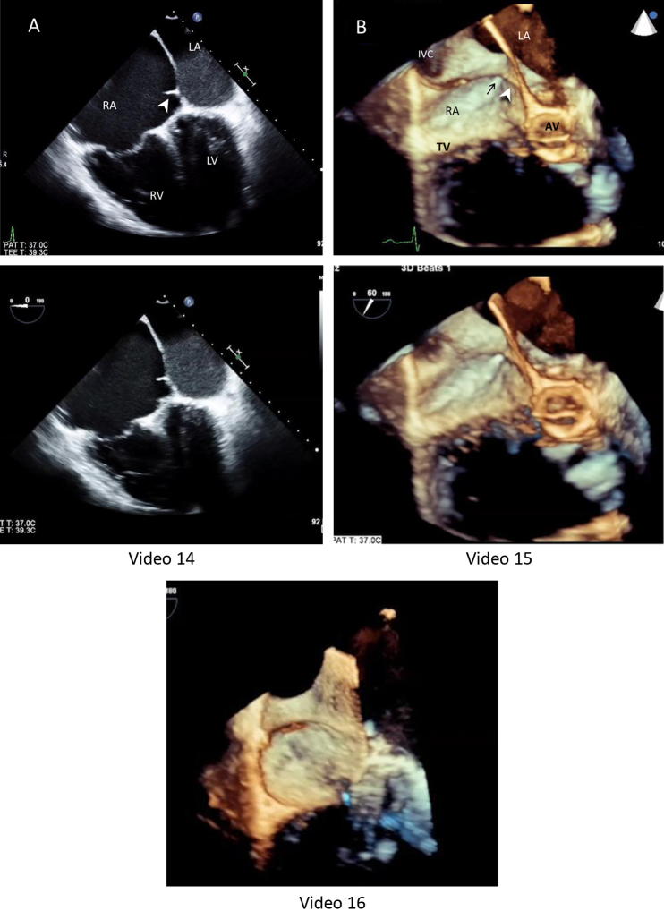
Case 5. Two-dimensional TEE images (A) show the ff-CTD membrane (arrowhead) attached to the atrial septum adjacent to the fossa ovalis, producing a telltale Y sign when the atrial septum is imaged just below the aortic root but above the coronary sinus (low four-chamber view). (B) TEE 3D views of ff-CTD membrane (arrowhead) using a semi–en face view of the atrial septum and TV annulus to best show the defect; black arrow = coronary sinus. AV, Aortic valve LA, left atrium; LV, left ventricle.
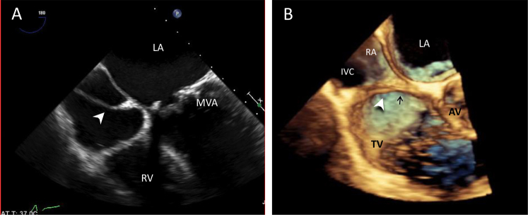
Case 6. Two-dimensional TEE image (A) shows the ff-CTD membrane (arrowhead) attached to the atrial septum adjacent to the fossa ovalis, producing a telltale Y sign when the atrial septum is imaged just below the aortic root but above the coronary sinus (low four-chamber view). (B) 3D TEE views of ff-CTD membrane (arrowhead) using a semi–en face view of the atrial septum and TV annulus to best show the defect; black arrow = coronary sinus. AV, Aortic valve LA, left atrium; MVA, MV annuloplasty ring.
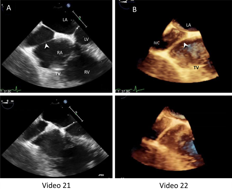
Case 7. Two-dimensional TEE images in the midesophageal four-chamber view. (A) The ff-CTD membrane (arrowhead) is attached to the atrial septum adjacent to the fossa ovalis, producing a telltale Y sign. (B) TEE in a bicaval view shows PFO closure device with precarious and incomplete deployment (arrow). LA, left atrium; LV, left ventricle.
Case 8. TEE images. (A) The ff-CTD membrane (arrowhead) attached to the atrial septum adjacent to the fossa ovalis, producing a telltale Y sign by 2D TEE. (B) TEE 3D view of the ff-CTD membrane (arrowhead) using a semi–en face view of the atrial septum and TV annulus to best show the defect. LA, Left atrium; LV, left ventricle.
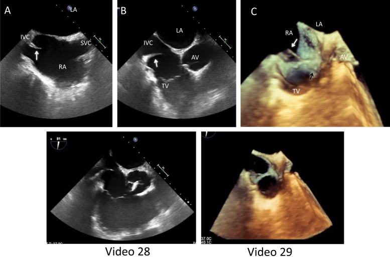
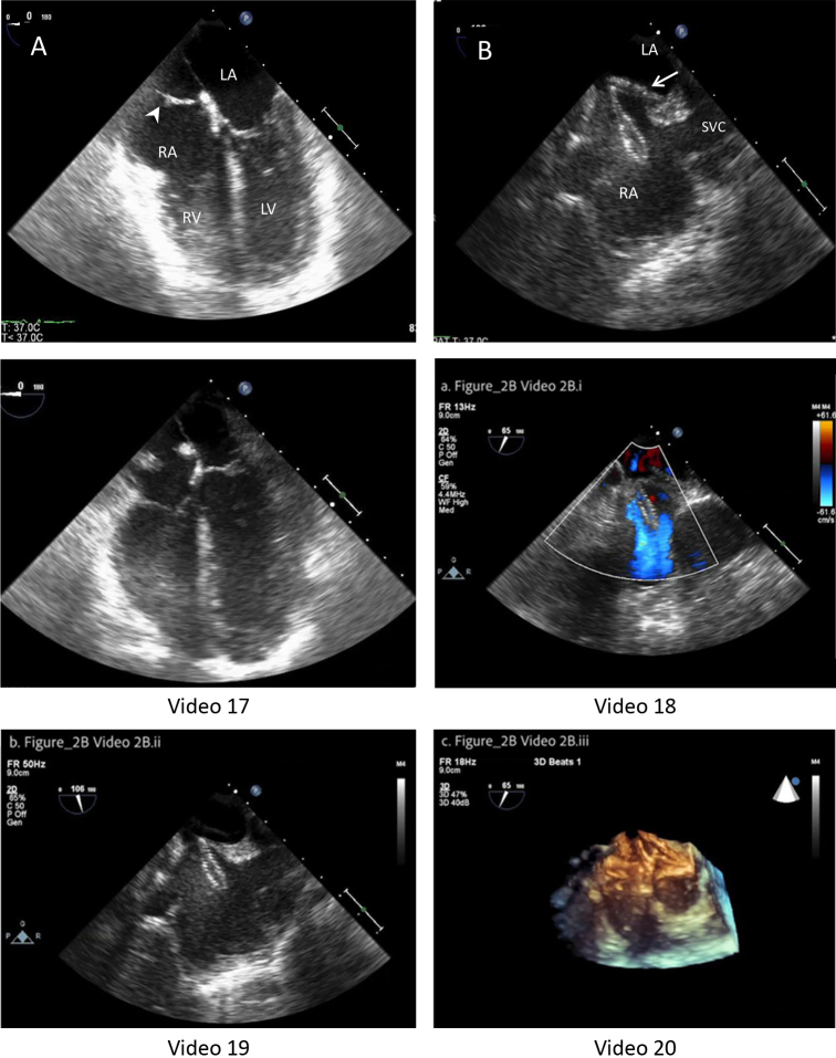
Case 9. Prominent EV (white arrows). (A) 2D TEE in midesophageal bicaval view, showing the IVC, SVC, RA, left atrium (LA), TV, and aortic valve (AV). (B) 2D TEE modified short-axis view of AV angulated toward the IVC and TV to show prominent EV (arrow), which terminates before the atrial septum. (C) 3D TEE image showing prominent EV, Eustachian ridge (black arrow), and coronary sinus. The Eustachian ridge extends to the inferior margin of the interatrial septum; the EV does not.
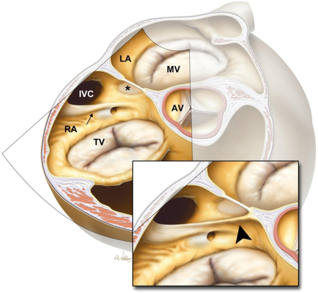
Illustration of modified 3D TEE reconstruction cut plane that is not a “standard” direct en face view of the interatrial septum but a modified view to better illustrate the relationship between the atrial septum and fossa ovalis (*) of either a prominent EV (small arrow) or the ff-CTD membrane illustrated in the inserted drawing (arrowhead), which extends onto the atrial septum and creates the Y sign on planar images. AV, Aortic valve; LA, left atrium.
Similar articles
-
Cor triatriatum dexter in an adult diagnosed by transesophageal echocardiography: a case report.J Am Soc Echocardiogr. 1995 Nov-Dec;8(6):952-7. doi: 10.1016/s0894-7317(05)80025-2. J Am Soc Echocardiogr. 1995. PMID: 8611301
-
Cor triatriatum dexter, atrial septal defect, and Ebstein's anomaly in an adult given a diagnosis by transthoracic and transesophageal echocardiography: a case report.J Am Soc Echocardiogr. 2004 Jul;17(7):780-2. doi: 10.1016/j.echo.2004.03.036. J Am Soc Echocardiogr. 2004. PMID: 15220906
-
Cor triatriatum dexter associated with atrial septal defect: Management in a complex clinical case.Echocardiography. 2017 Nov;34(11):1725-1729. doi: 10.1111/echo.13714. Echocardiography. 2017. PMID: 29178296
-
[Echocardiographic evaluation in unoperated congenital heart disease in adults].Herz. 1999 Jun;24(4):276-92. doi: 10.1007/BF03043879. Herz. 1999. PMID: 10444707 Review. German.
-
[Echocardiography in cor triatriatum dexter].Rev Port Cardiol. 1993 Dec;12(12):1043-8, 1001. Rev Port Cardiol. 1993. PMID: 8117458 Review. Portuguese.
Cited by
-
Percutaneous Edge-to-Edge Tricuspid Valve Repair in a Patient with Cor Triatriatum Dexter: A Case Report.J Cardiovasc Dev Dis. 2021 Sep 14;8(9):111. doi: 10.3390/jcdd8090111. J Cardiovasc Dev Dis. 2021. PMID: 34564129 Free PMC article.
-
Asymptomatic cor triatriatum dexter detected decades after cardiac surgery.J Med Ultrason (2001). 2025 Jul;52(3):323-324. doi: 10.1007/s10396-025-01532-5. Epub 2025 Apr 20. J Med Ultrason (2001). 2025. PMID: 40253672 No abstract available.
-
Mysterious Infantile Cyanosis: An Imaging Case Series.CASE (Phila). 2021 Aug 20;5(5):267-272. doi: 10.1016/j.case.2021.07.013. eCollection 2021 Oct. CASE (Phila). 2021. PMID: 34712868 Free PMC article.
-
Ridges and Pouches: A Case Series of Anomalous Atrial Septal Fusion.CASE (Phila). 2019 Dec 4;4(1):7-17. doi: 10.1016/j.case.2019.10.009. eCollection 2020 Feb. CASE (Phila). 2019. PMID: 32099937 Free PMC article.
-
Cor Triatriatum Dexter With a Sinus Venosus Atrial Septal Defect in a 50-Year-Old Woman: A Case Report.Cureus. 2024 Feb 2;16(2):e53477. doi: 10.7759/cureus.53477. eCollection 2024 Feb. Cureus. 2024. PMID: 38439997 Free PMC article.
References
-
- Hansing C.E., Young W.P., Rowe G.G. Cor triatriatum dexter. Persistent right sinus venosus valve. Am J Cardiol. 1972;30:559–564. - PubMed
-
- Krasemann Z., Scheld H.H., Tjan T.D., Krasemann T. Cor triatriatum: short review of the literature upon ten new cases. Herz. 2007;32:506–510. - PubMed
-
- Hoye D.J., Wilson E.C., Fyfe D.A., Guzzetta N.A. Cor triatriatum dexter: a rare cause of neonatal cyanosis. Anesth Analg. 2010;110:716–718. discussion 718. - PubMed
-
- Sarikouch S., Blanz U., Sandica E., Beerbaum P. Adult congenital heart disease: cor triatriatum dextrum. J Thorac Cardiovasc Surg. 2006;132:164–165. - PubMed
Publication types
LinkOut - more resources
Full Text Sources
