

The diversity of lobula plate tangential cells (LPTCs) in the Drosophila motion vision system
- PMID: 31709462
- PMCID: PMC7073280
- DOI: 10.1007/s00359-019-01380-y
The diversity of lobula plate tangential cells (LPTCs) in the Drosophila motion vision system
Abstract
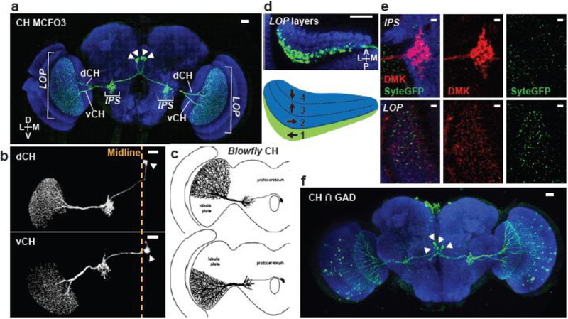
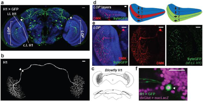
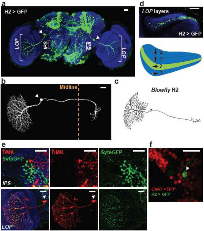
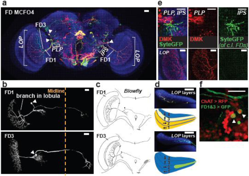
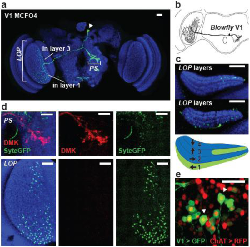
To navigate through the environment, animals rely on visual feedback to control their movements relative to their surroundings. In dipteran flies, visual feedback is provided by the wide-field motion-sensitive neurons in the visual system called lobula plate tangential cells (LPTCs). Understanding the role of LPTCs in fly behaviors can address many fundamental questions on how sensory circuits guide behaviors. The blowfly was estimated to have ~ 60 LPTCs, but only a few have been identified in Drosophila. We conducted a Gal4 driver screen and identified five LPTC subtypes in Drosophila, based on their morphological characteristics: LPTCs have large arborizations in the lobula plate and project to the central brain. We compared their morphologies to the blowfly LPTCs and named them after the most similar blowfly cells: CH, H1, H2, FD1 and FD3, and V1. We further characterized their pre- and post-synaptic organizations, as well as their neurotransmitter profiles. These anatomical features largely agree with the anatomy and function of their likely blowfly counterparts. Nevertheless, several anatomical details indicate the Drosophila LPTCs may have more complex functions. Our characterization of these five LPTCs in Drosophila will facilitate further functional studies to understand their roles in the visual circuits that instruct fly behaviors.
Keywords: Drosophila; LPTC; Lobula plate; Motion vision; Optomotor.
Conflict of interest statement
Figures
Similar articles
-
Synaptic organization of lobula plate tangential cells in Drosophila: Dalpha7 cholinergic receptors.J Neurogenet. 2009;23(1-2):200-9. doi: 10.1080/01677060802471684. J Neurogenet. 2009. PMID: 19306209
-
Columnar cells necessary for motion responses of wide-field visual interneurons in Drosophila.J Comp Physiol A Neuroethol Sens Neural Behav Physiol. 2012 May;198(5):389-95. doi: 10.1007/s00359-012-0716-3. Epub 2012 Mar 13. J Comp Physiol A Neuroethol Sens Neural Behav Physiol. 2012. PMID: 22411431 Free PMC article.
-
Neurons forming optic glomeruli compute figure-ground discriminations in Drosophila.J Neurosci. 2015 May 13;35(19):7587-99. doi: 10.1523/JNEUROSCI.0652-15.2015. J Neurosci. 2015. PMID: 25972183 Free PMC article.
-
How fly neurons compute the direction of visual motion.J Comp Physiol A Neuroethol Sens Neural Behav Physiol. 2020 Mar;206(2):109-124. doi: 10.1007/s00359-019-01375-9. Epub 2019 Nov 5. J Comp Physiol A Neuroethol Sens Neural Behav Physiol. 2020. PMID: 31691093 Free PMC article. Review.
-
Neuromodulation of insect motion vision.J Comp Physiol A Neuroethol Sens Neural Behav Physiol. 2020 Mar;206(2):125-137. doi: 10.1007/s00359-019-01383-9. Epub 2019 Dec 6. J Comp Physiol A Neuroethol Sens Neural Behav Physiol. 2020. PMID: 31811398 Review.
Cited by
-
Populations of local direction-selective cells encode global motion patterns generated by self-motion.Sci Adv. 2022 Jan 21;8(3):eabi7112. doi: 10.1126/sciadv.abi7112. Epub 2022 Jan 19. Sci Adv. 2022. PMID: 35044821 Free PMC article.
-
Building a circuit through correlated spontaneous neuronal activity in the developing vertebrate and invertebrate visual systems.Genes Dev. 2021 May 1;35(9-10):677-691. doi: 10.1101/gad.348241.121. Epub 2021 Apr 22. Genes Dev. 2021. PMID: 33888564 Free PMC article. Review.
-
Neural mechanisms to incorporate visual counterevidence in self-movement estimation.Curr Biol. 2023 Nov 20;33(22):4960-4979.e7. doi: 10.1016/j.cub.2023.10.011. Epub 2023 Nov 1. Curr Biol. 2023. PMID: 37918398 Free PMC article.
-
A Bio-Inspired Visual Neural Model for Robustly and Steadily Detecting Motion Directions of Translating Objects Against Variable Contrast in the Figure-Ground and Noise Interference.Biomimetics (Basel). 2025 Jan 14;10(1):51. doi: 10.3390/biomimetics10010051. Biomimetics (Basel). 2025. PMID: 39851767 Free PMC article.
-
Visual processing in the fly, from photoreceptors to behavior.Genetics. 2023 May 26;224(2):iyad064. doi: 10.1093/genetics/iyad064. Genetics. 2023. PMID: 37128740 Free PMC article.
References
Publication types
MeSH terms
Substances
Grants and funding
LinkOut - more resources
Full Text Sources
Molecular Biology Databases
