. 2020 Jan;9(1):302-312.
doi: 10.1002/cam4.2689. Epub 2019 Nov 10.

Anti-tumor effects of an antagonistic mAb against the ASCT2 amino acid transporter on KRAS-mutated human colorectal cancer cells

Affiliations

Anti-tumor effects of an antagonistic mAb against the ASCT2 amino acid transporter on KRAS-mutated human colorectal cancer cells

Yuta Hara et al. Cancer Med. 2020 Jan.

Abstract

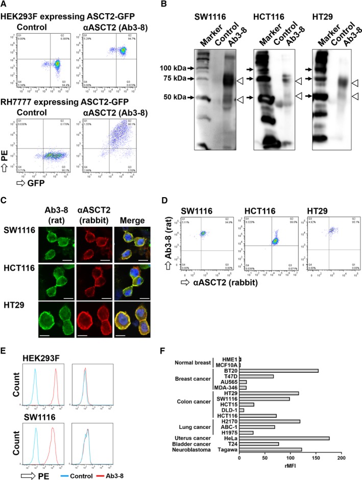
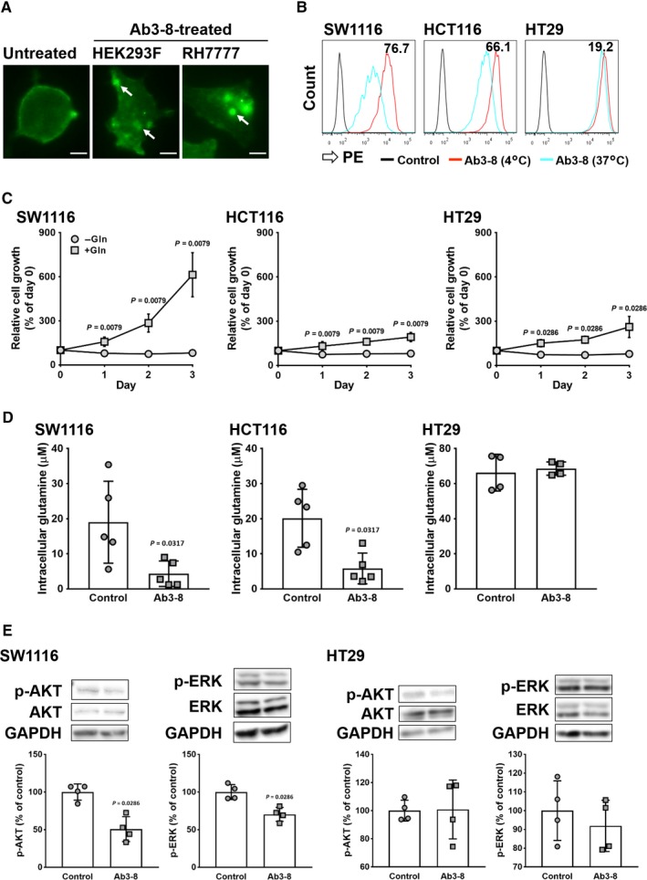
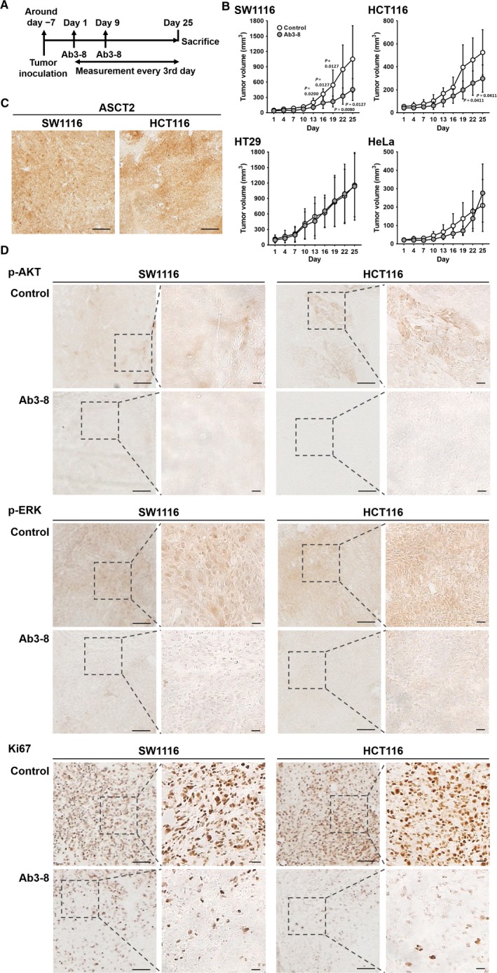
KRAS mutations are detected in numerous human cancers, but there are few effective drugs for KRAS-mutated cancers. Transporters for amino acids and glucose are highly expressed on cancer cells, possibly to maintain rapid cell growth and metabolism. Alanine-serine-cysteine transporter 2 (ASCT2) is a primary transporter for glutamine in cancer cells. In this study, we developed a novel monoclonal antibody (mAb) recognizing the extracellular domain of human ASCT2, and investigated whether ASCT2 can be a therapeutic target for KRAS-mutated cancers. Rats were immunized with RH7777 rat hepatoma cells expressing human ASCT2 fused to green fluorescent protein (GFP). Splenocytes from the immunized rats were fused with P3X63Ag8.653 mouse myeloma cells, and selected and cloned hybridoma cells secreting Ab3-8 mAb were established. This mAb reacted with RH7777 transfectants expressing ASCT2-GFP proteins in a GFP intensity-dependent manner. Ab3-8 reacted with various human cancer cells, but not with non-cancer breast epithelial cells or ASCT2-knocked out HEK293 and SW1116 cells. In SW1116 and HCT116 human colon cancer cells with KRAS mutations, treatment with Ab3-8 reduced intracellular glutamine transport, phosphorylation of AKT and ERK, and inhibited in vivo tumor growth of these cells in athymic mice. Inhibition of in vivo tumor growth by Ab3-8 was not observed in HT29 colon and HeLa uterus cancer cells with wild-type KRAS. These results suggest that ASCT2 is an excellent therapeutic target for KRAS-mutated cancers.

Keywords: ASCT2; CRC; KRAS; glutamine; mAb.


Conflict of interest statement

The authors declare no potential conflicts of interest.

Figures

Figure 1
Figure 1
Specificity of the Ab3‐8 anti‐human ASCT2 rat mAb. A, FCM analysis of Ab3‐8 against HEK293 (upper) and RH7777 (lower) cells expressing ASCT2‐GFP. PE, phycoerythrin. B, Immunoprecipitation. Lysates of SW1116, HCT116 and HT29 cells were immunoprecipitated with Ab3‐8, and then detected using a commercial anti‐ASCT2 rabbit mAb. Arrowhead, position of human ASCT2 proteins. C, PFA‐fixed cells were stained with a combination of Ab3‐8 and anti‐ASCT2 rabbit mAb, followed by Alexa Fluor 488‐conjugated anti‐rat IgG and Alexa Fluor 647‐conjugated anti‐rabbit IgG. Nuclei were counterstained with 4′,6‐diamidino‐2‐phenylindole (DAPI). Images were taken at 60 × magnification. Scale bar, 10 μm. D, PFA‐fixed cells were stained with the same antibodies as described above. E, ASCT2‐KO HEK293 and SW1116 cells were reacted with Ab3‐8, followed by PE‐conjugated anti‐rat IgG. The reactivity of Ab3‐8 with these cells was analyzed by flow cytometry. F, FCM analysis of cell surface expression of ASCT2 proteins in various human cell lines. The indicated cells were stained with Ab3‐8, followed by PE‐conjugated anti‐rat IgG. From values of the mean fluorescence intensity (MFI), the ratio (+mAb/ −mAb) of MFI (rMFI) was calculated.
Figure 2
Figure 2
Inhibitory effects of Ab3‐8 on ASCT2 functions. A, Microscopy study of ASCT2 internalization. HEK293 and RH7777 cells overexpressing ASCT2‐GFP were reacted with Ab3‐8 at 37°C for 1 h. Images were taken at 20 × magnification. Arrows indicate internalized ASCT proteins. Scale bar, 10 μm. B, Quantitative analysis of ASCT2 internalization using FCM. Human CRC cells were incubated with Ab3‐8 at 4°C or 37°C for 1 h, followed by PE‐conjugated secondary antibody. Numeric data in each panel indicate internalization (%) calculated from the reactivity (MFI) of Ab3‐8 with CRC cells at 37°C and 4°C. C, Glutamine‐dependent cell growth. Cells were cultured with or without glutamine (2 mmol/L) for 3 d. Relative cell growth was measured every 24 h. Each dot represents the mean of four (HT29) or five (SW1116 and HCT116) independent experiments. D, Intracellular glutamine uptake. Cells were incubated in PBS in the presence of Ab3‐8 (30 μg/mL) for 2 h, and then treated with 2 mmol/L glutamine for 10 min. Each dot represents value of individual experiments. E,Cells were incubated in serum‐ and glutamine‐free media in the presence of Ab3‐8 for 24 h, and then treated with 2 mmol/L glutamine for 15 min. Total and phosphorylated (p‐) AKT and ERK protein levels were measured by western blotting. GAPDH was used as a loading control. Each dot represents value of individual experiments. Data are expressed as mean ± SD of at least four independent experiments
Figure 3
Figure 3
Anti‐tumor effects of Ab3‐8. Cells were injected subcutaneously into the left/right flanks of male nude mice. A, Time course of the in vivo anti‐tumor study. B, Tumor volumes of CRC and HeLa cells were measured every 3 d using digital calipers, and were quantified using the formula: volume [mm3] = (length [mm]) × (width [mm])2 × 0.5. Ab3‐8 (100 μg/mouse) was intraperitoneally administered on days 1 and 9. Each dot represents the mean of 4‐9 mice. Data are expressed as the mean ± SD. Tumor volume of individual mice shows in Figure S3. C,Tumor sections from control mice were subjected to immunoperoxidase staining using Ab3‐8. Scale bar, 20 μm. D, Tumor sections were subjected to immunoperoxidase staining using the indicated antibodies. Representative images of the SW1116‐ and HCT116‐derived tumor sections, which immune labeled with p‐AKT, p‐ERK and Ki67. Images were obtained at 4 × magnification. Scale bar, 20 μm. Insets, higher magnification of the boxes. Images were obtained at 20 × magnification. Scale bar, 20 μm

References

    1. Ferlay J, Soerjomataram I, Dikshit R, et al. Cancer incidence and mortality worldwide: sources, methods and major patterns in GLOBOCAN 2012. Int J Cancer. 2015;136(5):E359‐386. - PubMed
    1. Arnold M, Sierra MS, Laversanne M, Soerjomataram I, Jemal A, Bray F. Global patterns and trends in CRC cancer incidence and mortality. Gut. 2017;66(4):683‐691. - PubMed
    1. Normanno N, Tejpar S, Morgillo F, De Luca A, Van Cutsem E, Ciardiello F. Implications for KRAS status and EGFR‐targeted therapies in metastatic CRC. Nat Rev Clin Oncol. 2009;6(9):519‐527. - PubMed
    1. Irahara N, Baba Y, Nosho K, et al. NRAS mutations are rare in colorectal cancer. Diagn Mol Pathol. 2010;19(3):157‐163. - PMC - PubMed
    1. Chang Y‐Y, Lin P‐C, Lin H‐H, et al. Mutation spectra of RAS gene family in colorectal cancer. Am J Surg. 2016;212(3):537‐544. - PubMed

Publication types

MeSH terms