. 2019 Dec 20;39(12):BSR20192414.
doi: 10.1042/BSR20192414.

Endoplasmic reticulum Ca2+ release causes Rieske iron-sulfur protein-mediated mitochondrial ROS generation in pulmonary artery smooth muscle cells

Affiliations

Endoplasmic reticulum Ca2+ release causes Rieske iron-sulfur protein-mediated mitochondrial ROS generation in pulmonary artery smooth muscle cells

Dapeng Dong et al. Biosci Rep. .

Retraction in

Abstract

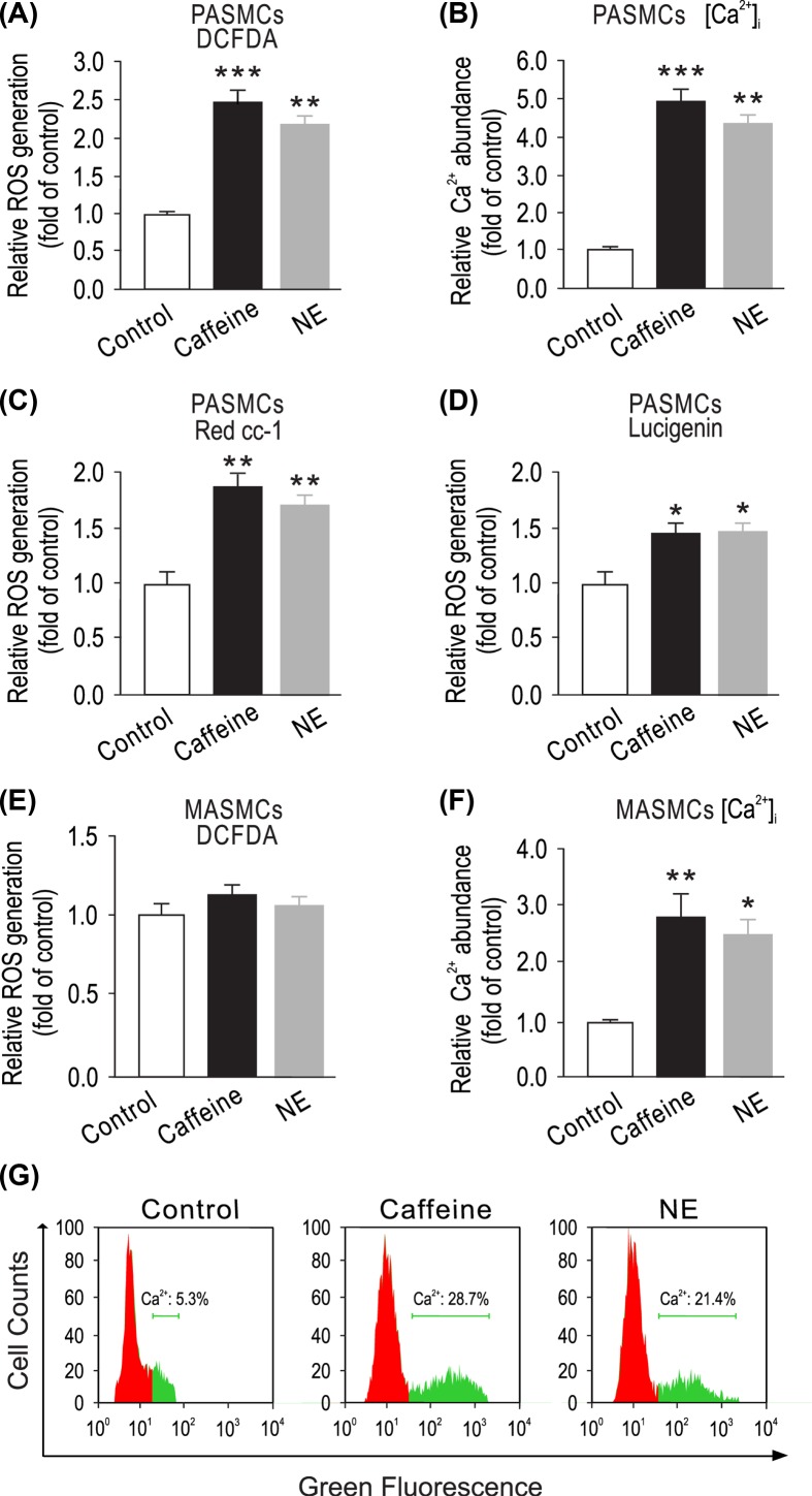
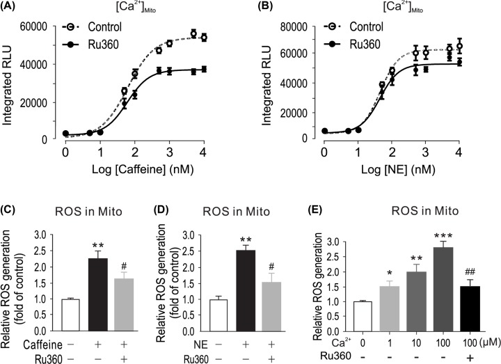
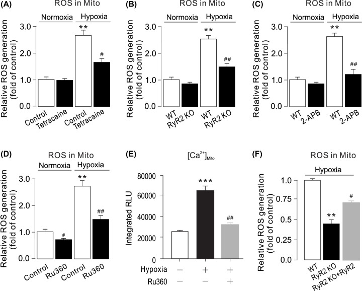
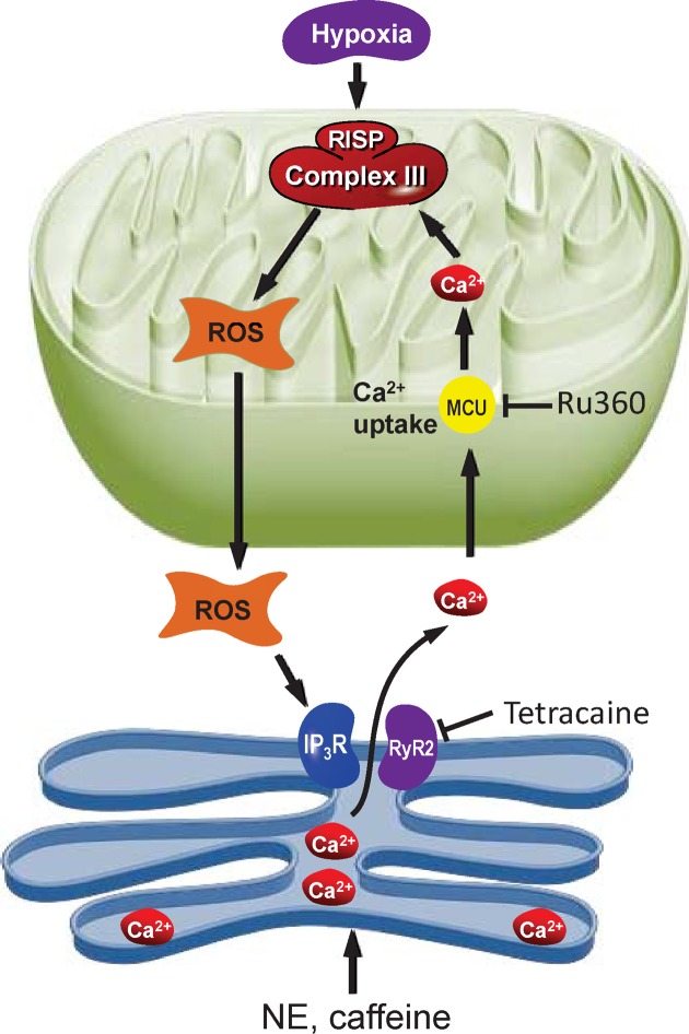
Mitochondrial reactive oxygen species (ROS) cause Ca2+ release from the endoplasmic reticulum (ER) via ryanodine receptors (RyRs) in pulmonary artery smooth muscle cells (PASMCs), playing an essential role in hypoxic pulmonary vasoconstriction (HPV). Here we tested a novel hypothesis that hypoxia-induced RyR-mediated Ca2+ release may, in turn, promote mitochondrial ROS generation contributing to hypoxic cellular responses in PASMCs. Our data reveal that application of caffeine to elevate intracellular Ca2+ concentration ([Ca2+]i) by activating RyRs results in a significant increase in ROS production in cytosol and mitochondria of PASMCs. Norepinephrine to increase [Ca2+]i due to the opening of inositol 1,4,5-triphosphate receptors (IP3Rs) produces similar effects. Exogenous Ca2+ significantly increases mitochondrial-derived ROS generation as well. Ru360 also inhibits the hypoxic ROS production. The RyR antagonist tetracaine or RyR2 gene knockout (KO) suppresses hypoxia-induced responses as well. Inhibition of mitochondrial Ca2+ uptake with Ru360 eliminates N- and Ca2+-induced responses. RISP KD abolishes the hypoxia-induced ROS production in mitochondria of PASMCs. Rieske iron-sulfur protein (RISP) gene knockdown (KD) blocks caffeine- or NE-induced ROS production. Taken together, these findings have further demonstrated that ER Ca2+ release causes mitochondrial Ca2+ uptake and RISP-mediated ROS production; this novel local ER/mitochondrion communication-elicited, Ca2+-mediated, RISP-dependent ROS production may play a significant role in hypoxic cellular responses in PASMCs.

Keywords: Intracellular calcium; Ryanodine receptor; mitochondrial ROS.


Conflict of interest statement

The authors declare that there are no competing interests associated with the manuscript.

Figures

Figure 1
Figure 1. Elevation of [Ca2+]i by caffeine or norepinephrine increases ROS production in cytosol of PASMCs, but not MASMCs
(A) PASMCs were treated with caffeine (200 μM) or norepinephrine (20 μM) for 5 min. Intracellular ROS generation was measured using DCFDA assay. (B) PASMCs were exposed to caffeine (200 μM) or norepinephrine (20 μM) for 5 min, and Ca2+ levels were then detected using a Ca2+ quantification kit. (C) Red CC-1 and lucigenin (D) assay was used to determine ROS generation in PASMCs. (E) MASMCs were treated with caffeine (200 μM) or norepinephrine (20 μM) for 5 min. Intracellular ROS generation was measured using DCFDA assay. (F) MASMCs were exposed to caffeine (200 μM) or norepinephrine (20 μM) for 5 min, and Ca2+ levels were then detected using a Ca2+ quantification kit. Data represent mean ± SEM; n = 6, *P < 0.05; **P < 0.01; ***P < 0.001, compared with control by two-tails Student’s t test. (G) Fluo3-AM staining of cytosolic Ca2+ ions in PASMCs. PASMCs were treated with caffeine (200 μM) or norepinephrine (20 μM) for 5 min, then stained with 0.5 μ M Fluo-3-AM in HBSS buffer.
Figure 2
Figure 2. Caffeine or norepinephrine elevates mitochondrial ROS by mitochondrial Ca2+ influx ([Ca2+]mito)
(A) [Ca2+]mito was determined by mitochondria-targeted double-mutated aequorin (pcDNA3.1+/mit-2mutAEQ), which was described in ‘Materials and Methods’ section. Mitochondria Ca2+ uniporter inhibitor Ru360 (1 μM) decreased [Ca2+]mito caused by caffeine or norepinephrine (B and C); PASMCs were treated with caffeine (200 μM) or norepinephrine (20 μM) (D) for 5 min. Mitochondrial ROS generation was measured using a Mitochondrial ROS Detection Assay Kit. (E) Freshly isolated mitochondria from PASMCs were exposed with different concentrations of Ca2+ in the presence or absence of Ru360 (1 μM); mitochondria ROS were determined by DCFDA assay. Data were obtained from three separate experiments. *P < 0.05; **P < 0.01; ***P < 0.001 compared with Ca2+ (0 μM) group. #P<0.05, ##P < 0.01 compared Ca2+ (100 μM) with absence of Ru360 group.
Figure 3
Figure 3. Inhibition or genetic deletion of RyR2 blocks hypoxic ROS production in PASMCs
(A) Intracellular ROS production was detected by DCFDA assay in PASMCs treated with RyRs inhibitor tetracaine (1 μM) for 5 min followed by hypoxia for 5 min. (B) Intracellular Ca2+ levels were then detected using a Ca2+ quantification kit. Data were obtained from three separate experiments. *P < 0.05; **P < 0.01; ***P < 0.001 compared with normoxic control group, and #P < 0.05; ##P < 0.01 compared with hypoxic control group. (C) Western blots of RyR2 expression in WT and RyR2 KO PASMCs. (D) PASMCs from wild-type (WT) and RyR2 KO mice were exposed to normoxia or hypoxia for 5 min. ROS were measured in cells by DCFDA assay. (E) Intracellular Ca2+ levels were then detected using a Ca2+ quantification kit. Data were obtained from three separate experiments. **P < 0.01 compared with normoxic WT group, and ##P < 0.01 compared with hypoxic WT group. (F) Mitochondrial ROS generation upon NE treatment in WT and RyR2 KO PASMCs.
Figure 4
Figure 4. Inhibition or genetic deletion of RyR2 blocks hypoxic ROS production in mitochondria of PASMCs
(A) PASMCs were pretreated with tetracaine (1 μM) (C) for 5 min followed by hypoxia for 5 min. Mitochondrial ROS generation was measured using a Mitochondrial ROS Detection Assay Kit. **P < 0.01 compared with normoxia control group, and ##P < 0.01 compared with hypoxia control group. (B) PASMCs from WT and RyR2 KO mice were exposed to normoxia or hypoxia for 5 min. Mitochondrial ROS were measured by a Mitochondrial ROS Detection Assay Kit. Data were obtained from three separate experiments. **P < 0.01 compared with control, and #P < 0.05; ##P < 0.01 compared with WT group. (C) WT and RyR2 KO PASMCs were pretreated with 2-APB (20 μM) for 5 min followed by hypoxia. Mitochondrial ROS generation were measured using a Mitochondrial ROS Detection Assay Kit. **P < 0.01 compared with normoxia control group, and ##P < 0.01 compared with hypoxia control group group. (D) Cells were treated with Ru360 (1 μM) for 5 min, then exposed to caffeine (20 mM) for 5 min. mitochondrial ROS were measured by using a Mitochondrial ROS Detection Assay Kit. **P < 0.01; ***P < 0.001 compared with normoxia control group, and ##P < 0.01 compared with hypoxia control group. [Ca2+]mito was determined by mitochondria-targeted double-mutated aequorin (pcDNA3.1+/mit-2mutAEQ). Ru360 (1 μM) decreased hypoxia-caused [Ca2+]mito. (E) RyR2 KO PASMCs were transfected with RyR2 overexpression plasmids. All groups were treated with hypoxia for 5 min. Mitochondrial ROS generation were measured using a Mitochondrial ROS Detection Assay Kit. **P < 0.01 compared with WT group, and #P < 0.05 compared with WT RyR2 KO group.
Figure 5
Figure 5. RISP gene knockdown blocks hypoxia-, caffeine- or NE- induced ROS production in PASMCs
(A) Western blots of RISP expression in PASMCs uninfected, infected with lentiviral RISP shRNA, and non-silencing (NS) shRNA. (B) Mitochondrial and Cytosolic ROS (C) were determined after hypoxia treatment. RISP knockdown inhibited hypoxia-induced ROS increase. Data were obtained from three separate experiments. **P < 0.01 compared with normoxia NS_shRNA group, and #P < 0.05; ##P < 0.01 compared with hypoxia NS_shRNA groups. (D) RISP KO PASMCs were transfected with RISP overexpression plasmids. All groups were treated with hypoxia for 5 min. Mitochondrial ROS generation were measured using a Mitochondrial ROS Detection Assay Kit. **P < 0.01 compared with WT group, and #P < 0.05 compared with WT RISP KO group. (E) PASMCs were treated with caffeine (200 μM) (E andF) or NE (20 μM) (G and H) for 5 min in non-infected, infected with non-silencing (NS) shRNA PASMCs or lentiviral encoding RISP shRNA. Mitochondria and Cytosolic ROS were determined after treatment. Data were obtained from three separate experiments. ***P < 0.001 compared with non-infected control group, and #P < 0.05; ##P < 0.01 compared with caffeine-treated NS_shRNA group.
Figure 6
Figure 6. ROS and Calcium crosstalk between endoplasmic reticulum and mitochondria
The ER is a major site of calcium ions (Ca2+) storage within muscle cells. Calcium from ER cisternae is flowing mainly through calcium release channels as inositol 1,4,5-trisphosphate receptors (IP3R) and ryanodine receptors (RyR). High levels of calcium stimulate respiratory chain activity leading to higher amounts of reactive oxygen species (ROS) through RISP. ROS can further target ER-based calcium channels leading to increased release of calcium and further increased ROS levels.

Similar articles

Cited by

References

    1. Moudgil R., Michelakis E.D. and Archer S.L. (2005) Hypoxic pulmonary vasoconstriction. J. Appl. Physiol. 98, 390–403, (1985) 10.1152/japplphysiol.00733.2004 - DOI - PubMed
    1. Sylvester J.T., Shimoda L.A., Aaronson P.I. and Ward J.P. (2012) Hypoxic pulmonary vasoconstriction. Physiol. Rev. 92, 367–520 10.1152/physrev.00041.2010 - DOI - PMC - PubMed
    1. Rowan S.C., Keane M.P., Gaine S. and McLoughlin P. (2016) Hypoxic pulmonary hypertension in chronic lung diseases: novel vasoconstrictor pathways. Lancet Respir. Med. 4, 225–236 10.1016/S2213-2600(15)00517-2 - DOI - PubMed
    1. Perez-Vizcaino F., Cogolludo A. and Moreno L. (2010) Reactive oxygen species signaling in pulmonary vascular smooth muscle. Respir. Physiol. Neurobiol. 174, 212–220 10.1016/j.resp.2010.08.009 - DOI - PubMed
    1. Du W., Frazier M., McMahon T.J. and Eu J.P. (2005) Redox activation of intracellular calcium release channels (ryanodine receptors) in the sustained phase of hypoxia-induced pulmonary vasoconstriction. Chest 128, 556S–855S 10.1378/chest.128.6_suppl.556S - DOI - PubMed

Publication types

MeSH terms