. 2020 Feb;117(2):362-371.
doi: 10.1002/bit.27220. Epub 2019 Nov 28.

"Trim"ming PolyQ proteins with engineered PML

Affiliations

"Trim"ming PolyQ proteins with engineered PML

Neha Dhar et al. Biotechnol Bioeng. 2020 Feb.

Abstract

Protein abnormalities are the major cause of neurodegenerative diseases such as spinocerebellar ataxia (SCA). Protein misfolding and impaired degradation leads to the build-up of protein aggregates inside the cell, which may further cause cellular degeneration. Reducing levels of either the soluble misfolded form of the protein or its precipitated aggregate, even marginally, could significantly improve cellular health. Despite numerous pre-existing strategies to target these protein aggregates, there is considerable room to improve their specificity and efficiency. In this study, we demonstrated the enhanced intracellular degradation of both monomers and aggregates of mutant ataxin1 (Atxn1 82Q) by engineering an E3 ubiquitin ligase enzyme, promyelocytic leukemia protein (PML). Specifically, we showed enhanced degradation of both soluble and aggregated Atxn1 82Q in mammalian cells by targeting this protein using PML fused to single chain variable fragments (scFvs) specific for monomers and aggregates of the target protein. The ability to solubilize Atxn1 82Q aggregates was due to the PML-mediated enhanced SUMOylation of the target protein. This ability to reduce the intracellular levels of both misfolded forms of Atxn1 82Q may not only be useful for treating SCA, but also applicable for the treatment of other PolyQ disorders.

Keywords: PML; aggregate; degradation; monomer; spinocerebellar ataxia.


Figures

Figure 1.
Figure 1.. Characterization of the influence of PML-scFv fusion proteins on levels of Atxn1 82Q-GFP.
A) Characterization of levels of Atxn1 82Q-GFP in HEK 293T cells using flow cytometry 24 hours after co-transfection with plasmids encoding Atxn1 82Q-GFP and vector control/ PML/PML-scFv fusion proteins. GFP intensity was measured as median fluorescence intensity. RMFI= MFI (SAMPLE)/MFI (VECTOR CONTROL). Values represent mean ± sd, n=3. B) Characterization of levels of Atxn1 82Q-GFP by flow cytometry, 48 hrs after co-transfection with plasmids encoding Atxn1 82Q-GFP and vector control/ PML/PML-scFv fusion proteins. Values represent mean ± sd, n=3. C) Characterization of levels of Atxn1 82Q-GFP in HEK 293T cells by Western Blotting. HEK 293T cells were co-transfected with Atxn1 82Q-GFP and 1-VC, 2-PML, 3- PML ΔSRS2,4-PML-6E, 5-PML-MW1, 6-PML-FRZ. For total cell lysate, cells were processed with 2% SDS buffer and probed by western blotting with an anti-GFP antibody (1:500). For fractionation, cells were processed in NP 40 buffer and the soluble fraction in this buffer was labelled as NS. Pellet was dissolved in SDS buffer, resulting in the SS fraction. VC: Vector Control.
Figure 2.
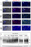
Figure 2.. Solubilization and SUMOylation by PML/PML-scFv fusion proteins.
A) HEK 293T cells were co-transfected with Atxn1 82Q-GFP and VC/PML/PML-scFvs (PML-MW1, PML-6E, PML-FRZ) in four-chambered glass bottom cover-glass. Cells were processed for DAPI staining and incubated with PML-specific mouse antibody, followed by further incubation with a Cy3-conjugated-Anti-mouse secondary antibody. A Spinning-Disc Confocal microscope was used to acquire images at 40×. Left column shows the DAPI staining overlaid with Bright field image in order to show nuclear staining. The images in the middle column represent an overlay of DAPI and Red channel, where Cy3-stained PML/PML-scFvs are seen as red specks localized around the nucleus (marked using yellow arrows in the middle column). The images in the right column represent an overlay of DAPI, Red, and Green channels, where along with PML/PML-scFv expression, Atxn1 82Q-GFP is seen either as intense green dots (Red arrow) for cells co-transfected with VC/ PML-FRZ (First and 5th row), as partially diffuse green dots for cells expressing PML (2nd row) and almost completely solubilized Atxn1 82Q-GFP for cells expressing PML-MW1 (4th row). Images were processed using Volocity software. B) HEK 293T cells were co-transfected with Atxn1 82Q-GFP and VC/PML/PML-scFvs and after an incubation of 24 hours, total cell lysate was probed using anti-SUMO 2/3 antibodies in western blotting. The experiment was performed with three biological repeats for each construct, as grouped by the marked lines. The samples were run on two separate gels as shown by a line between the samples but both blots were imaged together. Sumo 2/3 conjugates appear as a smear above anticipated Atxn1 82Q-GFP band at 122 KDa. VC: Vector Control.
Figure 3.
Figure 3.. Aggregation kinetics of Atxn1 82Q-GFP.
HEK 293T cells were co-transfected with Atxn1 82Q-GFP and VC/ PML/ PMLΔSRS2/PML-scFv fusion proteins. After 24 hours of incubation, cells were sorted and cells with Atxn1 82Q-GFP aggregates were counted using an epifluorescence microscope. The graph shows the fraction of HEK 293T cells without visible Atxn1 82Q-GFP aggregates as a function of time (hours). Vector control (green circle); PML (Red square); PMLΔSRS2 (brown triangle); PML-6E (inverted purple triangle); PML-MW1 (orange diamond); and PML-FRZ (black circle). The values represent mean ± sd, n=3. VC: Vector Control.
Figure 4:
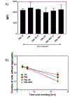
Figure 4:. Characterization of the influence of PML-scFv fusion proteins in the presence of pre-formed Atxn1 82Q-GFP aggregates.
A) HEK 293T cells were co-transfected with Atxn1 82Q-GFP and PML/PML-6E/MW1 encoded in Doxycycline inducible pTRE3G plasmid. PML/PML-scFv expression was induced by Doxycycline treatment, 48 hours after co-transfection in half of the wells for each construct. The remaining uninduced wells formed the control groups, denoted as UI. Total Atxn1 82Q-GFP present after 24 hours of Doxycycline treatment was measured as MFI using flow cytometry. B) Aggregation kinetics were studied for cells expressing pre-formed aggregates of Atxn1 82Q-GFP co-transfected with VC, PML, or PML-6E/MW1. As explained for (A), after an incubation of 48 hours, cells were induced with Doxycycline. Twenty four hours after Doxycycline induction, cells were sorted and the number of cells with visible protein aggregates were counted for the next 24 hours. The fraction of cells without aggregates was plotted against time post-seeding. The first data point is at 3 hours post seeding. Both plots represent mean ± sd, n=3
Figure 5.
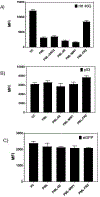
Figure 5.. Characterizing specificity of PML-scFvs.
HEK293T cells were co-transfected with VC/ PML/PML-MW1/PML-6E/FRZ and (A) Htt 46Q-GFP, (B) p53-GFP, and (C) eGFP. Twenty-four hours after co-transfection, cells were trypsinized and processed for flow-cytometric analysis. Target protein level was measure as MFI. All three plots represent mean± sd, n=3 VC: Vector Control.

References

    1. Black LR, & Aiken C (2010). TRIM5alpha disrupts the structure of assembled HIV-1 capsid complexes in vitro. J Virol, 84(13), 6564–6569. doi:10.1128/jvi.00210-10 - DOI - PMC - PubMed
    1. Caussinus E, Kanca O, & Affolter M (2011). Fluorescent fusion protein knockout mediated by anti-GFP nanobody. Nat Struct Mol Biol, 19(1), 117–121. doi:10.1038/nsmb.2180 - DOI - PubMed
    1. Dantuma NP, & Bott LC (2014). The ubiquitin-proteasome system in neurodegenerative diseases: precipitating factor, yet part of the solution. Front Mol Neurosci, 7, 70. doi:10.3389/fnmol.2014.00070 - DOI - PMC - PubMed
    1. De Genst E, Messer A, & Dobson CM (2014). Antibodies and protein misfolding: From structural research tools to therapeutic strategies. Biochim Biophys Acta, 1844(11), 1907–1919. doi:10.1016/j.bbapap.2014.08.016 - DOI - PubMed
    1. Dobson CM (2003). Protein folding and misfolding. Nature, 426(6968), 884–890. doi:10.1038/nature02261 - DOI - PubMed

Publication types

MeSH terms

LinkOut - more resources