Review
. 2020 Mar 1;116(4):787-805.
doi: 10.1093/cvr/cvz301.

Pathophysiology and diagnosis of coronary microvascular dysfunction in ST-elevation myocardial infarction

Affiliations
Review

Pathophysiology and diagnosis of coronary microvascular dysfunction in ST-elevation myocardial infarction

Lara S F Konijnenberg et al. Cardiovasc Res. .

Abstract

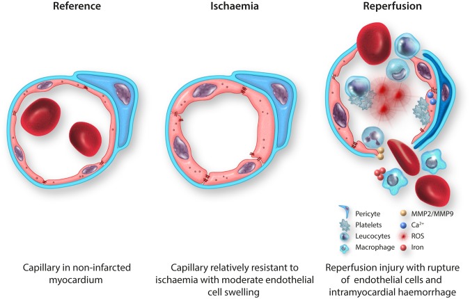
Early mechanical reperfusion of the epicardial coronary artery by primary percutaneous coronary intervention (PCI) is the guideline-recommended treatment for ST-elevation myocardial infarction (STEMI). Successful restoration of epicardial coronary blood flow can be achieved in over 95% of PCI procedures. However, despite angiographically complete epicardial coronary artery patency, in about half of the patients perfusion to the distal coronary microvasculature is not fully restored, which is associated with increased morbidity and mortality. The exact pathophysiological mechanism of post-ischaemic coronary microvascular dysfunction (CMD) is still debated. Therefore, the current review discusses invasive and non-invasive techniques for the diagnosis and quantification of CMD in STEMI in the clinical setting as well as results from experimental in vitro and in vivo models focusing on ischaemic-, reperfusion-, and inflammatory damage to the coronary microvascular endothelial cells. Finally, we discuss future opportunities to prevent or treat CMD in STEMI patients.

Keywords: Coronary microvascular dysfunction; Coronary microvascular endothelial cells; Intramyocardial haemorrhage; Microvascular reperfusion injury; ST-elevation myocardial infarction.


Figures

Graphical Abstract
Graphical Abstract
Figure 1
Figure 1
Relationship between CMR-defined microvascular obstruction, infarct size, and major adverse cardiac events. IS, infarct size; IS%LV, infarct size as percentage of left ventricle; LV, left ventricle; MACE, major adverse cardiac events; MO, CMR-defined microvascular obstruction. Values are Kaplan–Meier estimates in patients with IS%LV ≥25% vs. <25%, grouped by the presence or absence of MO, indicating the time to MACE during 2 years of follow-up. MACE is defined as a composite of cardiac death, myocardial reinfarction, and new congestive heart failure at 2 years of follow-up. Reprinted with permission from Van Kranenburg et al.
Figure 2
Figure 2
Intracoronary physiology: invasive measurements for coronary microvascular function after ST-elevation myocardial infarction. (A) Schematic overview of Pa, Pd, Tmn, APV, BR, CFR, IMR, HMR, RRR, IHDVPS, and Pzf. (B) With an intracoronary pressure and/or Doppler flow wire the following indices can be derived: Pa (red), Pd (green), BR, hyperaemic (dark blue lines) and rest (light blue lines) Tmn, hyperaemic (dark blue box) and rest (light blue box) Doppler APV, IHDVPS (pink dotted line) and Pzf (yellow dot). The right box shows the pressure–flow velocity loops and the calculation of the linear relationship between pressure and flow velocity over the mid-late phases of diastole (coloured in black). The higher the values of IHDVPS (the β term in the equation y = a + βx, where y = flow velocity and x = intracoronary pressure), the better the conductance of the microcirculation. Pzf is calculated from the intercept of the regression line with the pressure axis. (C) Formulas for CFR, IMR, HMR, RRR, IHDVPS, and Pzf. The arrow indicates higher or lower values for that formula when coronary microvascular dysfunction is present. Evidence on the ability of IHDVPS to reflect coronary microvascular dysfunction is controversial. See text for details. APV, average peak velocity; BR, basal resistance, CFR, coronary flow reserve; HMR, hyperaemic microvascular resistance; IHDVPS, instantaneous hyperaemic diastolic flow velocity–pressure slope; IMR, index of microcirculatory resistance; Pa, aortic pressure; Pd, distal pressure; Pzf, pressure at zero flow velocity; RRR, resistive reserve ratio; Tmn, mean transit time.
Figure 3
Figure 3
CMR images of a patient after acute anterior myocardial infarction. Typical example of a patient after acute anterior myocardial infarction with microvascular injury, demonstrating hyperintense oedematous myocardium on the T2-weighted image (A, V) compared to remote non-infarcted myocardium (O), with the corresponding delayed contrast-enhanced image showing the infarcted hyperenhanced infarcted myocardium (B, V) with a hypointense infarct core (asterisk) with microvascular injury. The area with microvascular injury has low T1 on the non-contrast T1 map (C, asterisk) and low T2*, whereas the area of infarction has increased T1 relaxation times and normal T2* decay times, not containing haemoglobin breakdown products. LV, left ventricle; RV, right ventricle.
Figure 4
Figure 4
Coronary microvascular endothelial cell dysfunction in reperfused STEMI. (A) TEM image of a capillary in non-infarcted myocardium (healthy reference) of a rat with thin endothelium, preserved interendothelial cell junctions and numerous pinocytotic vesicles (small arrow), surrounded by a basement membrane; 17 500×. (B) Schematic overview of a healthy reference capillary with an attached pericyte. (C) TEM image of a capillary in permanently ischaemic myocardium of a rat with a preserved endothelial cell lining and some localized endothelial cell swelling, surrounded by a basement membrane; 24 500×. (D) Schematic overview of a capillary in permanently ischaemic myocardium with some diffuse endothelial cell swelling and some destabilization of interendothelial cell junctions. (E) TEM image of a completely ruptured capillary—beyond the phase of endothelial cell swelling with blebs—in reperfused myocardium of a rat with ruptured endothelial cell lining (small arrows) and intramyocardial haemorrhage; 5800×. (F) Schematic overview of completely ruptured capillary in reperfused myocardium with massive production of ROS, elevated levels of cytosolic calcium activating the contractile elements of endothelial cells, platelet adhesion and aggregation, destabilization of interendothelial cell junctions, intramyocardial haemorrhage, and numerous inflammatory cells. ▸,  endothelial cell; B, basement membrane; CM, cardiomyocyte; E, erythrocyte; J, interendothelial cell junctions; M, mitochondrion; MMP, matrix metalloprotease; N, nucleus; P, pericyte; ROS, reactive oxygen species; V, pinocytotic vesicle.

References

    1. Cohen M, Boiangiu C, Abidi M.. Therapy for ST-segment elevation myocardial infarction patients who present late or are ineligible for reperfusion therapy. J Am Coll Cardiol 2010;55:1895–1906. - PubMed
    1. Van Kranenburg MV, Magro M, Thiele H, DeWaha S, Eitel I, Cochet A, Cottin Y, Atar D, Buser P, Wu E, Lee D, Bodi V, Klug G, Metzler B, Delewi R, Bernhardt P, Rottbauer W, Boersma E, Zijlstra F, Geuns RJ.. Prognostic value of microvascular obstruction and infarct size, as measured by CMR in STEMI patients. JACC Cardiovasc Imaging 2014;7:930–939. - PubMed
    1. Reffelmann T, Kloner RA.. Microvascular reperfusion injury: rapid expansion of anatomic no reflow during reperfusion in the rabbit. Am J Physiol Circ Physiol 2002;283:H1099–H1107. - PubMed
    1. Lima JAC, Wu KC, Becker LC, Melin JA, Gerber BL, Rochitte CE, Bluemke DA, McVeigh ER.. Microvascular obstruction and left ventricular remodeling early after acute myocardial infarction. Circulation 2012;101:2734–2741. - PubMed
    1. Kloner RA, Ganote CE, Jennings RB.. The ‘no reflow’ phenomenon after temporary coronary occlusion in the dog. J Clin Invest 1974;54:1496–1508. - PMC - PubMed

Publication types

MeSH terms