

Brainstem pathology in amyotrophic lateral sclerosis and primary lateral sclerosis: A longitudinal neuroimaging study
- PMID: 31711033
- PMCID: PMC6849418
- DOI: 10.1016/j.nicl.2019.102054
Brainstem pathology in amyotrophic lateral sclerosis and primary lateral sclerosis: A longitudinal neuroimaging study
Abstract
Background: Brainstem pathology is a hallmark feature of ALS, yet most imaging studies focus on cortical grey matter alterations and internal capsule white matter pathology. Brainstem imaging in ALS provides a unique opportunity to appraise descending motor tract degeneration and bulbar lower motor neuron involvement.
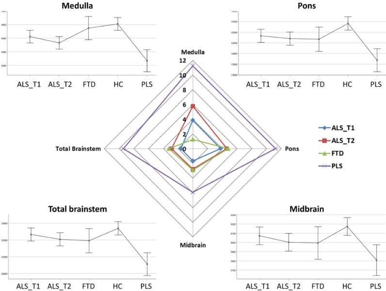
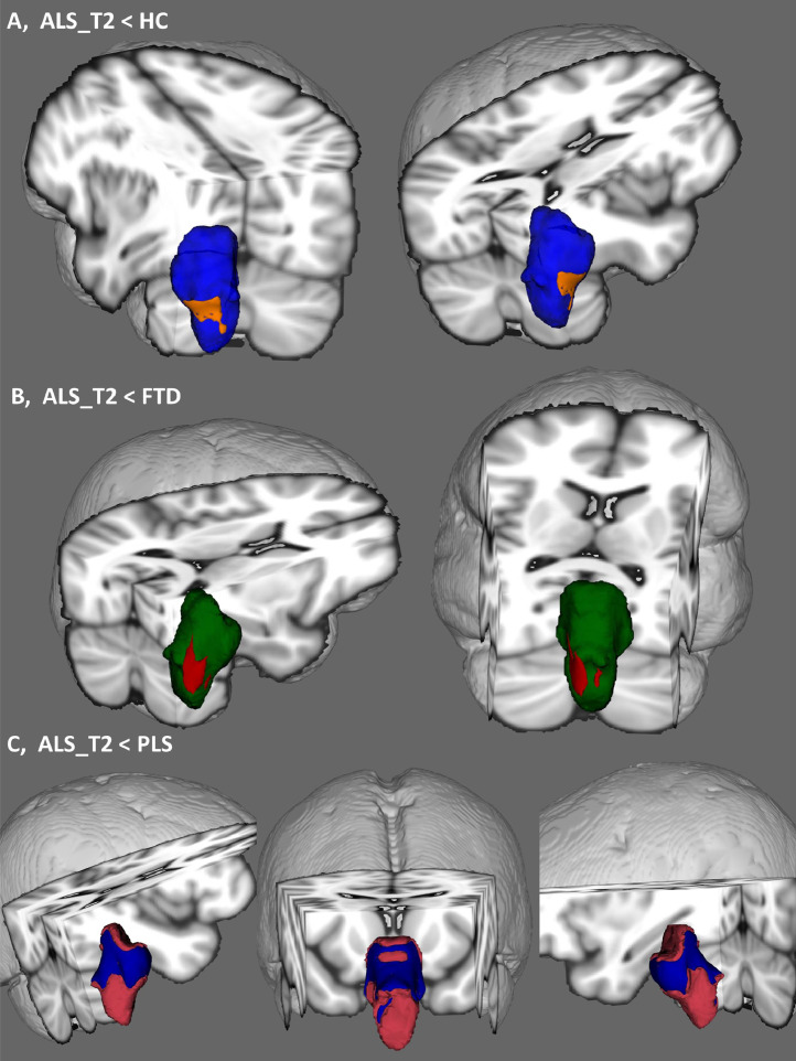
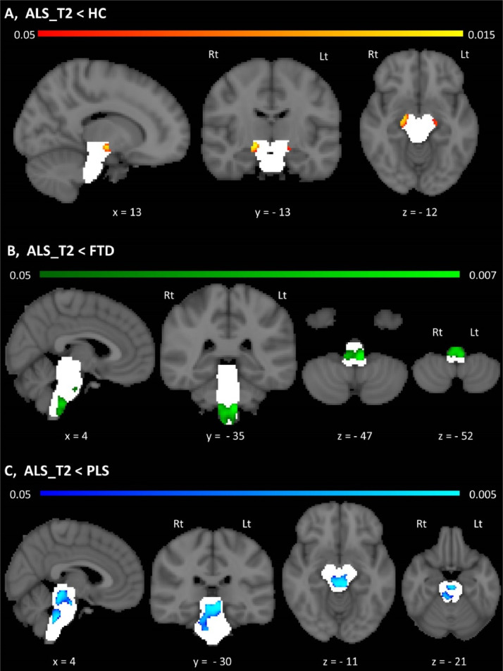
Methods: A prospective longitudinal imaging study has been undertaken with 100 patients with ALS, 33 patients with PLS, 30 patients with FTD and 100 healthy controls. Volumetric, vertex and morphometric analyses were conducted correcting for demographic factors to characterise disease-specific patterns of brainstem pathology. Using a Bayesian segmentation algorithm, the brainstem was segmented into the medulla, pons and mesencephalon to measure regional volume reductions, shape analyses were performed to ascertain the atrophy profile of each study group and region-of-interest morphometry was used to evaluate focal density alterations.
Results: ALS and PLS patients exhibit considerable brainstem atrophy compared to both disease- and healthy controls. Volume reductions in ALS and PLS are dominated by medulla oblongata pathology, but pontine atrophy can also be detected. In ALS, vertex analyses confirm the flattening of the medullary pyramids bilaterally in comparison to healthy controls and widespread pontine shape deformations in contrast to PLS. The ALS cohort exhibit bilateral density reductions in the mesencephalic crura in contrast to healthy controls, central pontine atrophy compared to disease controls, peri-aqueduct mesencephalic and posterior pontine changes in comparison to PLS patients. CONCLUS: ions: Computational brainstem imaging captures the degeneration of both white and grey matter components in ALS. Our longitudinal data indicate progressive brainstem atrophy over time, underlining the biomarker potential of quantitative brainstem measures in ALS. At a time when a multitude of clinical trials are underway worldwide, there is an unprecedented need for accurate biomarkers to monitor disease progression and detect response to therapy. Brainstem imaging is a promising addition to candidate biomarkers of ALS and PLS.
Keywords: Amyotrophic lateral sclerosis; Brainstem; Longitudinal study; Medulla; Mesencephalon; Motor neuron disease; Neuroimaging; Pons; Primary lateral sclerosis.
Copyright © 2019 The Author(s). Published by Elsevier Inc. All rights reserved.
Conflict of interest statement
Peter Bede is a member of the UK Motor Neuron Disease Association (MNDA) Research Advisory Panel, the steering committee of Neuroimaging Society in ALS (NiSALS) and the medical patron of the Irish Motor Neuron Disease Association (IMNDA). These affiliations had no impact on the design, analyses, and presentation of this work.
Figures
References
-
- Mitsumoto H., Brooks B.R., Silani V. Clinical trials in amyotrophic lateral sclerosis: why so many negative trials and how can trials be improved? Lancet Neurol. 2014;13(11):1127–1138. - PubMed
-
- Schuster C., Elamin M., Hardiman O. Presymptomatic and longitudinal neuroimaging in neurodegeneration–from snapshots to motion picture: a systematic review. J. Neurol. Neurosurg. Psychiatry. 2015;86(10):1089–1096. - PubMed
-
- Hardiman O., Doherty C.P., Elamin M. Springer International Publishing; Switzerland: 2016. Neurodegenerative Disorders: A Clinical Guide; pp. 1–336. 2016 ed. 2016, Springer Cham Heidelberg New York Dordrecht London©Springer International Publishing.
-
- Omer T., Finegan E., Hutchinson S., Doherty M., Vajda A., McLaughlin R.L., Pender N., Hardiman O., Bede P. Neuroimaging patterns along the ALS-FTD spectrum: a multiparametric imaging study. Amyotroph Lateral Scler Frontotemporal Degener. 2017;18(7-8):611–623. Epub 2017 May 31.PMID: 28562080. - PubMed
Publication types
MeSH terms
LinkOut - more resources
Full Text Sources
Medical
Miscellaneous
