. 2020 Mar 13;102(3):671-679.
doi: 10.1093/biolre/ioz209.

Transcript profiling of bovine embryos implicates specific transcription factors in the maternal-to-embryo transition

Affiliations

Transcript profiling of bovine embryos implicates specific transcription factors in the maternal-to-embryo transition

Yanina S Bogliotti et al. Biol Reprod. .

Abstract

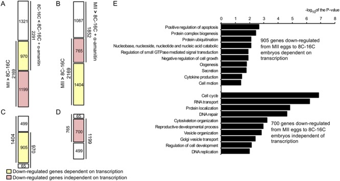
Full-grown oocytes are transcriptionally quiescent. Following maturation and fertilization, the early stages of embryonic development occur in the absence (or low levels) of transcription that results in a period of development relying on maternally derived products (e.g., mRNAs and proteins). Two critical steps occur during the transition from maternal to embryo control of development: maternal mRNA clearance and embryonic genome activation with an associated dramatic reprogramming of gene expression required for further development. By combining an RNA polymerase II inhibitor with RNA sequencing, we were able not only to distinguish maternally derived from embryonic transcripts in bovine preimplantation embryos but also to establish that embryonic gene activation is required for clearance of maternal mRNAs as well as to identify putative transcription factors that are likely critical for early bovine development.

Keywords: cow; embryo genome activation; gene expression; maternal-to-embryo transition; microRNA; preimplantation embryo; transcription factor.


Figures

Figure 1
Figure 1
Global transcriptome assessment of MII eggs and 8-to-16-cell embryos. A) Experimental design to identify embryonic genes expressed during early bovine development. RNA sequencing was performed using pools of 10 oocytes/embryos (3–4 replicates per group). B) Principal component analysis was done for all developmental stages with their replicates (MII, n = 4; 8-to-16-cell, n = 3; and 8-to-16-cell+alpha-amanitin, n = 4). Each point represents a replicate. Developmental stages were clearly separated from each other and replicates within stages were grouped together. C) Venn diagram comparing all expressed genes (RPKM > 0.4) between MII eggs and 8-to-16-cell embryos. 10 601 and 10 608 genes were found expressed in MII eggs and 8-to-16-cell embryos, respectively, representing ~ 39% of the total number of known genes. D) Gene ontology analysis of genes expressed in MII eggs and 8-to-16-cell embryos. Transcripts detected only in MII eggs (n = 1,060) or 8-to-16-cell embryos (n = 1,067) were subjected to GO analysis. Bar plot shows the −log10 of the P-value of the selected GO terms for DAVID biological processes.
Figure 2
Figure 2
Analysis of differentially expressed genes across developmental stages. A) Diagram showing the number of differentially expressed genes (FDR < 0.01, FC > 2) in oocytes and embryos. Up-headed arrows indicate the number of transcripts with increased relative abundance between stages and down-headed arrows show decreased abundance. B) Venn diagram showing the comparison among relative abundance of transcripts that increased from MII to 8–16 cell stage ± alpha-amanitin and those whose relative abundance in 8–16-cell embryos were decreased when alpha-amanitin was included in the culture medium. Some 2459 transcripts were products of transcription from the embryonic genome, whereas 719 transcripts were exclusively of maternal origin. C) Gene ontology analysis of newly transcribed embryonic and maternally derived transcripts. GO analysis was done on the 2459 embryonic genes and 719 maternal genes. Bar plot shows the −log10 of the P-value of the selected GO terms for biological processes using DAVID.
Figure 3
Figure 3
Effect of transcriptional activity on transcript turnover during MET. A) Comparison between the 2169 down-regulated transcripts from MII to 8-to-16-cell stage with the 2291 up-regulated transcripts when the embryos were cultured in the presence of alpha-amanitin. Transcripts whose relative abundance decrease was linked to transcription (n = 970) are denoted in yellow, whereas transcripts whose relative abundance decrease was not linked to transcription are denoted in red (n = 1199). B) Comparison between the 2169 down-regulated transcripts between MII eggs and 8-to-16-cell stage with the 1852 down-regulated transcripts from MII eggs to 8-to-16-cell+alpha-amanitin embryos. Transcripts whose relative abundance was linked to transcription (n = 1404) are denoted in yellow, whereas transcripts whose relative abundance decrease was not linked to transcription are denoted in red (n = 765). C) Comparison between the transcripts found in A and B to be down-regulated only in the presence of transcription from the embryonic genome (970 and 1,404). D) Comparison between the transcripts found in A and B to be down-regulated independent of transcription from the embryonic genome (1,199 and 765). E) Gene ontology analysis of transcripts whose relative abundance decreased from MII to 8-to-16-cell stage was linked or not linked to transcription. GO analysis was performed on the 905 transcripts whose decrease relative abundance from MII to the 8–16-cell stage was linked to transcription and on the 700 transcripts whose decrease relative abundance from MII to the 8–16-cell stage was not linked to transcription. Bar plot shows the −log10 of the P-value of the selected GO terms for biological processes using DAVID.
<b>Figure</b> 4
Figure 4
Transcription factors with enriched target motifs in the 2459 embryonic gene list. A) Transcription factors that could bind to the 29 enriched motifs were examined in the 719 maternal and 2459 embryonic gene lists. RPKM was used to measure expression levels. Red, nonexpressed. Green, expressed. B) Target gene overlap of three members of the KLF gene family. KLF5, KLF9, and KLF4 transcription factors can target 1141, 503, and 342 genes in the 2459 early embryonic gene list, respectively. C) Quantitative RT-PCR validation of transcription factor expression during bovine EGA. KLF4, KLF5, GSC, and OTX2 were expressed in 8-cell embryos, and their expression was blocked by alpha-amanitin. DGCR8, whose expression is not up-regulated during EGA, was used as a control gene. a,b,c; different letters represent P < 0.05. x,y,z; different letters represent P < 0.1.

References

    1. Abe K, Yamamoto R, Franke V, Cao M, Suzuki Y, Suzuki MG, Vlahovicek K, Svoboda P, Schultz RM, Aoki F. The first murine zygotic transcription is promiscuous and uncoupled from splicing and 3′ processing. EMBO J 2015; 34:1523–1537. - PMC - PubMed
    1. Svoboda P, Franke V, Schultz RM. Sculpting the Transcriptome during the oocyte-to-embryo transition in mouse. Curr Top Dev Biol 2015; 113:305–349. - PubMed
    1. Braude P, Bolton V, Moore S. Human gene expression first occurs between the four- and eight-cell stages of preimplantation development. Nature 1988; 332:459–461. - PubMed
    1. Misirlioglu M, Page GP, Sagirkaya H, Kaya A, Parrish JJ, First NL, Memili E. Dynamics of global transcriptome in bovine matured oocytes and preimplantation embryos. Proc Natl Acad Sci U S A 2006; 103:18905–18910. - PMC - PubMed
    1. Telford NA, Watson AJ, Schultz GA. Transition from maternal to embryonic control in early mammalian development: A comparison of several species. Mol Reprod Dev 1990; 26:90–100. - PubMed

Publication types

Substances