

Three-dimensional kinematic change of hindfoot during full weightbearing in standing: an analysis using upright computed tomography and 3D-3D surface registration
- PMID: 31711523
- PMCID: PMC6849314
- DOI: 10.1186/s13018-019-1443-z
Three-dimensional kinematic change of hindfoot during full weightbearing in standing: an analysis using upright computed tomography and 3D-3D surface registration
Abstract
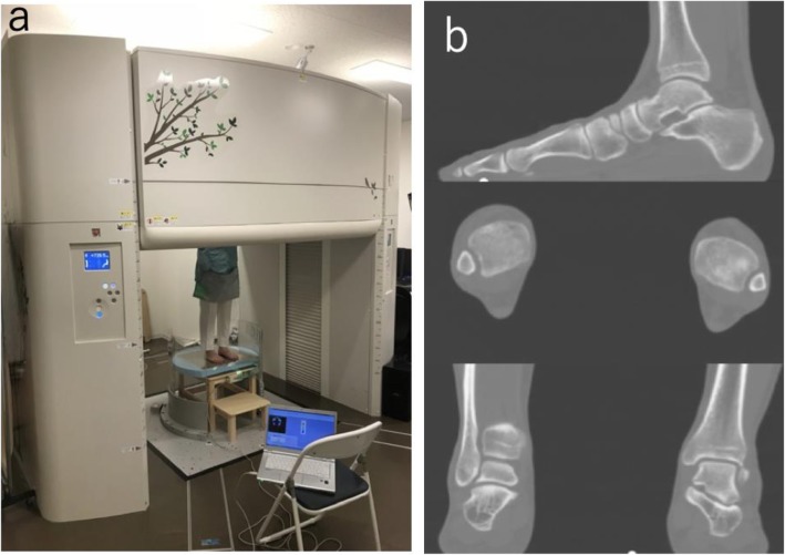
Background: Weightbearing of the hindfoot affects positional changes of the ankle joint and subtalar joint (ankle-joint complex [AJC]). However, it is difficult to assess the kinematic changes in the hindfoot in a natural full weightbearing condition using conventional CT or cone beam computed tomography (CT) due to limitations of acquiring foot images under a physiological weightbearing condition using those imaging modalities. Analysis of AJC kinematics using fluoroscopy and 2D-3D registration technique requires data on the number of steps and amount of time to build and match the bones. This study aimed to analyze the effect of full weightbearing on hindfoot motion when standing using upright CT and 3D-3D surface registration.
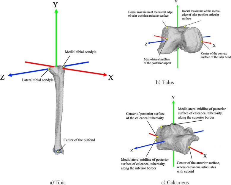
Methods: Forty-eight AJCs of 24 asymptomatic volunteers (13 women, 11 men) were examined under no weightbearing, 50% weightbearing, and single leg full weightbearing conditions while standing. The CT images were acquired from the distal femur to the whole foot using a 320-row upright CT scanner. The condition of each weightbearing stance was measured using a pressure mat. Bone-to-bone rotations of the talus relative to the tibia and calcaneus relative to the talus were evaluated using the surface registration technique. Image quality of the CT and intra- and interobserver reliabilities of the rotation angle were also evaluated.
Results: All CT images were excellent or good quality and the intra- and interobserver correlation coefficients for the angle were 0.996 and 0.995, respectively. The motion of the ankle joint and subtalar joint under 50% and 100% weightbearing were as follows (in degrees); the talus plantarflexed (5.1 ± 4.5 and 6.8 ± 4.8), inverted (1.3 ± 1.4 and 2.0 ± 1.6), and internally rotated (2.4 ± 4.2 and 4.3 ± 4.6) relative to the tibia, and the calcaneus dorsiflexed (2.8 ± 1.4 and 3.8 ± 1.7), everted (5.3 ± 2.6 and 8.0 ± 3.6), and externally rotated (3.0 ± 2.0 and 4.1 ± 2.4) relative to the talus, respectively.
Conclusions: The effect of weightbearing was clearly identified using an upright CT and the 3D-3D registration technique. Three-dimensional kinematics under static full weightbearing were opposite between the ankle and subtalar joints on their respective axes.
Keywords: Hindfoot; Surface registration; Upright computed tomography; Weightbearing.
Conflict of interest statement
The authors declare that they have no competing interests.
Figures


References
-
- Goto A, Moritomo H, Itohara T, Watanabe T, Sugamoto K. Three-dimensional in vivo kinematics of the subtalar joint during dorsi-plantarflexion and inversion-eversion. Foot Ankle Int. 2009;30(5):432–438. - PubMed
-
- Yamaguchi S, Sasho T, Kato H, Kuroyanagi Y, Banks SA. Ankle and subtalar kinematics during dorsiflexion-plantarflexion activities. Foot Ankle Int. 2009;30(4):361–366. - PubMed
-
- Kobayashi T, Saka M, Suzuki E, Yamazaki N, Suzukawa M, Akaike A, et al. In vivo kinematics of the talocrural and subtalar joints during weightbearing ankle rotation in chronic ankle instability. Foot Ankle Spec. 2014;7(1):13–19. - PubMed
MeSH terms
Grants and funding
LinkOut - more resources
Full Text Sources
Medical
