Review
. 2019 Dec;40(12):1006-1020.
doi: 10.1016/j.tips.2019.10.007. Epub 2019 Nov 8.

Targeting Muscarinic Acetylcholine Receptors for the Treatment of Psychiatric and Neurological Disorders

Affiliations
Review

Targeting Muscarinic Acetylcholine Receptors for the Treatment of Psychiatric and Neurological Disorders

Sean P Moran et al. Trends Pharmacol Sci. 2019 Dec.

Abstract

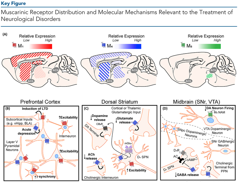
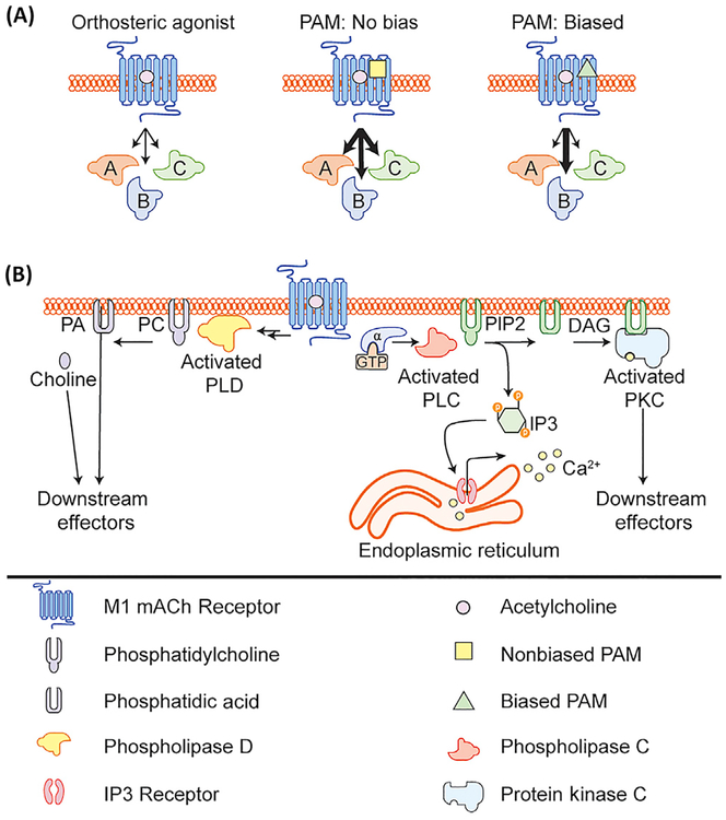
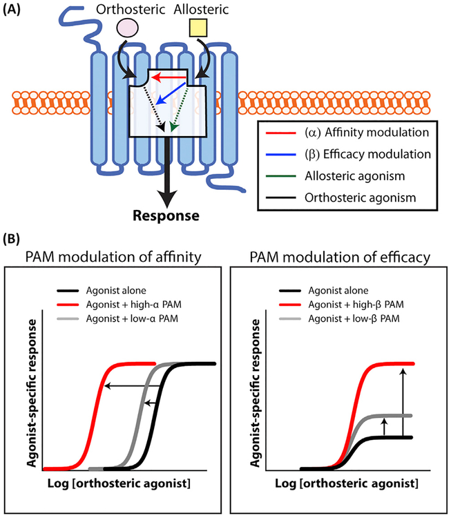
Muscarinic acetylcholine receptors (mAChR) play important roles in regulating complex behaviors such as cognition, movement, and reward, making them ideally situated as potential drug targets for the treatment of several brain disorders. Recent advances in the discovery of subtype-selective allosteric modulators for mAChRs has provided an unprecedented opportunity for highly specific modulation of signaling by individual mAChR subtypes in the brain. Recently, mAChR allosteric modulators have entered clinical development for Alzheimer's disease (AD) and schizophrenia, and have potential utility for other brain disorders. However, mAChR allosteric modulators can display a diverse array of pharmacological properties, and a more nuanced understanding of the mAChR will be necessary to best translate preclinical findings into successful clinical treatments.

Keywords: NAM; PAM; allosteric modulator; muscarinic acetylcholine receptor; neurological disorder; signal bias.


Figures

Figure 1.
Figure 1.
(A) Distribution of M1, M4, and M5 muscarinic acetylcholine (ACh) receptors in brain regions implicated in neurological dysfunction. The relative expression of each receptor subtype is indicated by its respective color gradient. M2 and M3 (not shown) muscarinic ACh receptors (mAChRs) are also expressed widely throughout the brain. M1 mAChRs are highly expressed in the cortex, hippocampus, and dorsal and ventral striatum, and are expressed at low levels in thalamic areas. M4 mAChRs are highly expressed in striatal regions, moderately expressed across the cortex and thalamus, and are poorly expressed in the hippocampus. M5 mAChR expression is restricted to the midbrain. Cholinergic projection neurons produce and release ACh from two distinct clusters – the basal forebrain nuclei (grey circle, left) which innervates cortical, hippocampal, and thalamic areas, and the brain stem nuclei (grey circle, right) which innervates midbrain, hindbrain, thalamic, and cerebellar areas. Cholinergic tone in the dorsal and ventral striatum is primarily provided by large cholinergic interneurons (not depicted). (B) In the prefrontal cortex, M1 mAChR activation induces a form of long-term depression (LTD) of glutamatergic inputs from subcortical areas including the ventral hippocampus (vHipp) and basolateral amygdala (BLA). M1 mAChR activation also increases the excitability of pyramidal neurons and GABAergic interneurons. Activation of M1 via interneurons can also increase gamma oscillation synchrony in the cortex. M4 mAChRs can acutely inhibit neurotransmitter release. (C) In the dorsal striatum, M4 mAChRs expressed on direct pathway D1 receptor-positive spiny projection neurons (SPNs) interact with metabotropic glutamate receptor 1 (mGlu1) to produce endocannabinoids which then bind to cannabinoid type 2 (CB2) receptors to inhibit local dopamine release. In addition, M4 activation can reduce both ACh release from local cholinergic interneurons and act as a heteroreceptor on glutamatergic terminals from the cortex and thalamus to reduce glutamate release. M1 mAChRs expressed on D1-SPNs increase the excitability of these neurons. (D) In the midbrain, cholinergic modulation of dopaminergic (DA) neurons in the ventral tegmental area (VTA) and direct pathway input into the substantia nigra reticulata (SNr) are relevant to neurological disorders. In the VTA (top), M5 mAChRs are expressed on VTA DA neurons and M5 negative allosteric modulators (NAMs) are hypothesized to reduce DA neuron firing. In the SNr (bottom), M4 mAChR activation on direct pathway D1-SPN terminals directly opposes increased GABA release mediated through D1-receptor activation by DA released from the substantia nigra pars compacta (SNpc, middle). M4 can also act as an autoreceptor and reduce ACh release from cholinergic projection terminals.
Figure 2.
Figure 2.. M1 Muscarinic Acetylcholine Receptor (mAChR) Allosteric Modulator-Induced Signal Bias.
(A) Schematic depicting the effects of acetylcholine (ACh) alone, ACh plus a non-biased M1 positive allosteric modulator (PAM), or ACh plus a biased M1 PAM on various downstream signaling cascades. (B) Activation of the M1 mAChR can lead to the activation of several downstream signaling pathways including canonical activation of Gaq signaling leading to activation of phospholipase C, release of calcium, and activation of PKC. M1 mAChR activation can also lead to activation of phospholipase D, through an unknown mechanism. Abbreviations: DAG, diacylglycerol; IP3, inositol trisphosphate; PA, phosphatidic acid; PC, phosphatidylcholine; PIP2, phosphatidylinositol bisphosphate.
Figure I.
Figure I.. Allosteric Modulator Modes of Action.
(A) Allosteric modulators (yellow squares) bind to a topographically and structurally distinct site on the muscarinic receptor to modulate orthosteric agonist (ACh, pink) affinity (red) and/or efficacy (blue). Binding of positive allosteric agonists (PAMs) or ago-PAMs can also directly induce receptor signaling in the absence of the orthosteric agonist (green). (B) (Left) Allosteric modulators that robustly modulate agonist affinity (high α value, red) will result in a large leftward shift in the orthosteric agonist concentration–response curve. By contrast, allosteric modulators that weakly enhance agonist affinity (low α value, grey) result in a modest leftward shift in the orthosteric agonist concentration–response curve. (Right) Allosteric modulators that strongly modulate agonist efficacy (high β value, red) may result in a large increase in the orthosteric agonist maximal response. By contrast, allosteric modulators that weakly enhance agonist efficacy (low β value, grey) result in a modest increase in the orthosteric agonist response. Sigmoidal curves were generated using Graphpad Prism8 (www.graphpad.com).

References

    1. Picciotto MR et al. (2012) Acetylcholine as a neuromodulator: cholinergic signaling shapes nervous system function and behavior. Neuron 76, 116–129 - PMC - PubMed
    1. Soreq H (2015) Checks and balances on cholinergic signaling in brain and body function. Trends Neurosci. 38, 448–458 - PubMed
    1. Sarter M et al. (2014) Deterministic functions of cortical acetylcholine. Eur. J. Neurosci 39, 1912–1920 - PMC - PubMed
    1. Hasselmo ME and Sarter M (2011) Modes and models of forebrain cholinergic neuromodulation of cognition. Neuropsychopharmacology 36, 52–73 - PMC - PubMed
    1. Wess J et al. (2007) Muscarinic acetylcholine receptors: mutant mice provide new insights for drug development. Nat. Rev. Drug Discov. 6, 721–733 - PubMed

Publication types

MeSH terms