Case Reports
. 2019 Nov 10;12(11):e231444.
doi: 10.1136/bcr-2019-231444.

Cancer-associated retinopathy in ampullary pancreatic cancer

Affiliations
Case Reports

Cancer-associated retinopathy in ampullary pancreatic cancer

Nima Ghadiri et al. BMJ Case Rep. .

Abstract

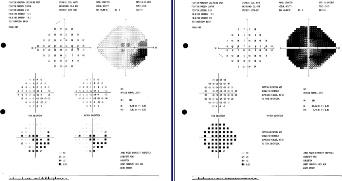
A 64-year-old woman presented with bilateral visual loss with shimmering photopsias as the only clinical manifestation of an occult pancreatic ampullary adenocarcinoma causing duct dilatation. Abnormal electroretinograms led to suspicion of cancer-associated retinopathy (CAR), and CT of the abdomen showed an underlying pancreatic malignancy, detected with subclinical liver function tests following diagnosis of CAR. Biopsy showed a T2N0M0 ampullary adenocarcinoma. The patient was managed with Whipple's procedure and adjuvant chemotherapy and has made a good recovery with no progression of her retinopathy. To our knowledge, this is one of the first descriptions of CAR in the context of pancreatic malignancy. It is atypical in its asymmetric presentation and favourable patient outcome. CAR is an important diagnosis to make, as ocular manifestations can be the only indication of an occult malignancy, resulting in a swifter diagnosis and potentially life-saving early intervention.

Keywords: ophthalmology; pancreatic cancer; retina.


Conflict of interest statement

Competing interests: None declared.

Figures

Figure 1
Figure 1
Paracentral scotoma in the right eye on visual field testing.
Figure 2
Figure 2
Fundus photography showing a normal fundus in the right (A) and left (B) eyes.
Figure 3
Figure 3
Optical coherence tomography findings showing the characteristic loss of photoreceptor inner segment–outer segment junctions as seen in autoimmune retinopathy in the right (A) and left (B) eyes.
Figure 4
Figure 4
Fundus autofluorescence showing a single focus of perivascular hypofluorescence on the left with no evidence of any hyperautofluorescence.
Figure 5
Figure 5
Abnormal ERGs: (A) pattern ERG demonstrating lack of response to stimulus even at a large field stimulus, (B) scotopic ERG showing diminished B-wave, and (C) photopic responses showing bilaterally delayed and small B-waves. ERG, electroretinogram.
Figure 6
Figure 6
CT of the abdomen demonstrating intrahepatic biliary duct dilatation (white arrows) and marked dilatation of the common bile duct (red arrow).

References

    1. Braithwaite T, Vugler A, Tufail A. Autoimmune retinopathy. Ophthalmologica 2012;228:131–42. 10.1159/000338240 - DOI - PubMed
    1. Weleber RG, Watzke RC, Shults WT, et al. . Clinical and electrophysiologic characterization of paraneoplastic and autoimmune retinopathies associated with Antienolase antibodies. Am J Ophthalmol 2005;139:780–94. 10.1016/j.ajo.2004.12.104 - DOI - PubMed
    1. Panzeri F, et al. Management of ampullary neoplasms: a tailored approach between endoscopy and surgery. World J Gastroenterol 2015;21:7970–87. 10.3748/wjg.v21.i26.7970 - DOI - PMC - PubMed
    1. Heckenlively JR, Ferreyra HA. Autoimmune retinopathy: a review and summary. Semin Immunopathol 2008;30:127–34. 10.1007/s00281-008-0114-7 - DOI - PubMed
    1. Grewal DS, Fishman GA, Jampol LM, et al. . Autoimmune retinopathy and antiretinal antibodies. Retina 2014;34:1023–41. 10.1097/01.iae.0000450880.26367.4e - DOI - PubMed

Publication types