. 2019 Nov 19;116(47):23714-23723.
doi: 10.1073/pnas.1906346116. Epub 2019 Nov 11.

Therapeutic targeting of tumor-associated myeloid cells synergizes with radiation therapy for glioblastoma

Affiliations

Therapeutic targeting of tumor-associated myeloid cells synergizes with radiation therapy for glioblastoma

Peng Zhang et al. Proc Natl Acad Sci U S A. .

Abstract

Tumor-associated myeloid cells (TAMCs) are key drivers of immunosuppression in the tumor microenvironment, which profoundly impedes the clinical response to immune-dependent and conventional therapeutic modalities. As a hallmark of glioblastoma (GBM), TAMCs are massively recruited to reach up to 50% of the brain tumor mass. Therefore, they have recently been recognized as an appealing therapeutic target to blunt immunosuppression in GBM with the hope of maximizing the clinical outcome of antitumor therapies. Here we report a nano-immunotherapy approach capable of actively targeting TAMCs in vivo. As we found that programmed death-ligand 1 (PD-L1) is highly expressed on glioma-associated TAMCs, we rationally designed a lipid nanoparticle (LNP) formulation surface-functionalized with an anti-PD-L1 therapeutic antibody (αPD-L1). We demonstrated that this system (αPD-L1-LNP) enabled effective and specific delivery of therapeutic payload to TAMCs. Specifically, encapsulation of dinaciclib, a cyclin-dependent kinase inhibitor, into PD-L1-targeted LNPs led to a robust depletion of TAMCs and an attenuation of their immunosuppressive functions. Importantly, the delivery efficiency of PD-L1-targeted LNPs was robustly enhanced in the context of radiation therapy (RT) owing to the RT-induced up-regulation of PD-L1 on glioma-infiltrating TAMCs. Accordingly, RT combined with our nano-immunotherapy led to dramatically extended survival of mice in 2 syngeneic glioma models, GL261 and CT2A. The high targeting efficiency of αPD-L1-LNP to human TAMCs from GBM patients further validated the clinical relevance. Thus, this study establishes a therapeutic approach with immense potential to improve the clinical response in the treatment of GBM and warrants a rapid translation into clinical practice.

Keywords: PD-L1; glioblastoma; immunotherapy; myeloid cell; radiotherapy.


Conflict of interest statement

Competing interest statement: There is a pending patent pertaining to the work presented in this manuscript.

Figures

Fig. 1.
Fig. 1.
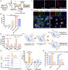
Engineering of therapeutic LNPs targeting glioma-associated TAMCs. (A) Schematic representation of nano-targeting of glioma-associated TAMCs. (CTL, cytotoxic T lymphocyte; Teff, effector T cell; PD-1, programmed cell death protein 1; IFNGR, IFN gamma receptor). (B and C) Flow cytometric quantification of PD-L1 expression among glioma-infiltrating immune cells in the GL261 glioma model, as determined by percentage of PD-L1+ cells (B) (blue, control; red, PE anti-mouse PD-L1), and MFI (C). Data are represented as mean ± SEM; n = 3; ***P < 0.001; determined by 1-way ANOVA with Tukey’s multiple comparisons test. (D) αPD-L1–functionalized LNP (αPD-L1-LNP) and naked LNP were characterized by cryo-EM, DLS, and zeta-potential. (Scale bar, 50 nm.)
Fig. 2.
Fig. 2.
αPD-L1-LNPs effectively target in-vitro–generated TAMC and impair PD-L1 recycling. (A) Schematic of in vitro generation of GL261 glioma-associated TAMCs. (B) Flow cytometric quantification of cellular binding of Rhod-PE–labeled LNPs in TAMCs after 1 h of binding at 4 °C. (C) Fluorescence microscopy images of cellular uptake of Rhod-PE–labeled LNPs by TAMCs after 1 h of incubation at 37 °C. (Scale bar, 50 µm.) (D) Intracellular trafficking of Rhod-PE–labeled αPD-L1-LNPs in TAMCs after 1 h of incubation at 37 °C. Cell membrane was stained by WGA, lysosome was stained by Lyso-Tracker DND26, and cell nucleus was stained by NucBlue. (Scale bar, 50 µm.) (E) Flow cytometric analysis of cellular uptake of Rhod-PE–labeled LNPs within a coculture of TAMCs and GL261 glioma cells after 1 and 4 h of incubation. (F) Schematic of PD-L1 internalization and recycling assay. (G and H) Flow cytometric analysis of cell-surface PD-L1 (G) and cell- surface–bound αPD-L1 (H). Cells were treated with unconjugated αPD-L1 or αPD-L1-LNP and collected after binding at 4 °C or subsequent incubation at 37 °C to allow internalization and recycling. PM was used as a recycling inhibitor. Data are represented as mean ± SEM; n = 3; *P < 0.05; ***P < 0.001; n.s., not significant; determined by 1-way ANOVA in B, G, and H or 2-way ANOVA in E with Tukey’s multiple comparisons test.
Fig. 3.
Fig. 3.
Therapeutic LNPs effectively impair viability and immunosuppressive activities of TAMCs. (A) Schematic of the chemical structure and LNP encapsulation of Dina. (B) Annexin V analysis of TAMCs 24 h after treatment of αPD-L1-LNP/Dina, αPD-L1-LNP, or Dina. (C) Expression of PD-L1 on TAMCs 24 h post stimulation with IFNγ, as determined by RT-qPCR. mRNA levels were normalized to beta-actin and reported relative to control TAMC expression. (D) Flow cytometric analysis of PD-L1 expression on TAMCs 24 h post stimulation with IFNγ, as presented by MFI. (E) Representative histograms of proliferating CD8+ T cells 72 h after being cocultured with nontreated TAMCs (blue) or 25 nM of αPD-L1-LNP/Dina–treated TAMCs (red), as traced by Cell Trace Violet and compared to CD8+ T cells alone (gray-shaded region). Data are represented as mean ± SEM; n = 3; *P < 0.05; ***P < 0.001; determined by 1-way ANOVA with Tukey’s multiple comparisons test.
Fig. 4.
Fig. 4.
Therapeutic LNPs actively target TAMCs in an ex vivo and an in vivo glioma model and extend survival of glioma-bearing mice. (A) Schematic of isolating immune infiltrates in GL261 glioma model. (B) Flow cytometric analysis of distribution of Rhod-PE–labeled LNPs among immune cell subsets, as represented by MFI (n = 3). (C and D) Flow cytometric analysis of glioma-associated immune cells after treatment with αPD-L1-LNP/Dina at a Dina concentration of 0, 25, and 50 nM for 72 h (n = 4). (C) Representative gating of CD45high CD11b TIL, CD45high CD11b+ TAMC, and CD45int CD11b+ microglia. (D) The cell abundance was determined by cell counts and flow cytometry analysis, as normalized to nontreated control. (E) Distribution of Rhod-PE–labeled αPD-L1-LNPs at a brain tumor site 24 h post injection. (Scale bar, 100 μm.) (F) Survival curves of GL261-bearing mice after 2 administrations of saline, drug-free αPD-L1-LNP, Iso-LNP/Dina, or αPD-L1-LNP/Dina at 2.5 mg/kg Dina on days 7 and 14 after intracranial implantation of 5 × 10⁴ GL261 glioma cells; n = 7–8 mice per group. Data are represented as mean ± SEM; *P < 0.05; **P < 0.01; ***P < 0.001; determined by 1-way ANOVA in D or 2-way ANOVA in B with Tukey’s multiple comparisons test or log-rank method with P values adjusted by Bonferroni correction in F.
Fig. 5.
Fig. 5.
Irradiation up-regulates PD-L1 expression on TAMCs and enhances targeted delivery to TAMCs. (A) RT-qPCR and flow cytometric quantification of PD-L1 expression on TAMCs as normalized to control TAMC expression. (B) Flow cytometric quantification of cellular uptake of Rhod-PE–labeled LNPs in TAMCs after 1 h of incubation, as presented by the percentage of NP+ cells (blue, nontreated TAMCs; red, NP-treated TAMCs). (C) Flow cytometric quantification of percentage of PD-L1–positive TAMCs (blue, Iso control; red, PE anti-PD-L1). (D) Cell circle analysis of TAMCs treated with phosphate-buffered saline, RT (8 Gy), αPD-L1-LNP, αPD-L1-LNP/Dina (25 nM Dina), or RT+αPD-L1-LNP/Dina (25 nM Dina). Data are represented as mean ± SEM; n = 3; *P < 0.05; ***P < 0.001; determined by Student’s t test in A or 1-way ANOVA with Tukey’s multiple comparisons test in D.
Fig. 6.
Fig. 6.
Therapeutic nanoparticles synergize with radiation therapy to eliminate TAMCs and improve therapeutic efficacy in glioma-bearing mice. (A) Schematic representation of the experimental workflow of combination therapy in GL261- or CT2A-bearing mice. Selected groups received RT (2 Gy×4) as monotherapy or combination therapy. (B) Survival curves of mice received intracranial implantation of 2 × 10⁵ GL261 glioma cells and 2 administrations of saline, drug-free αPD-L1-LNP or αPD-L1-LNP/Dina (5 mg/kg Dina). n = 10 mice per group. (CG) Flow cytometric analysis of GL261 glioma-associated immune cells. The abundance of TAMCs was determined by cell counts and flow cytometry analysis, as normalized to control mice (C). Subsets of TAMCs (M, M-MDSC; P, PMN-MDSC; T, TAM) were analyzed by abundance (D) and percentage (E). PD-L1 expression on TAMCs was determined by percentage of the PD-L1–positive population (F) and MFI (G). Data are represented as mean ± SEM; n = 3 to 4. (H) The experimental workflow of combination therapy through intranasal delivery. (I) Survival curves of mice received intracranial implantation of 5 × 10⁴ GL261 glioma cells and 8 administrations of saline or αPD-L1-LNP/Dina (5 mg/kg Dina) through an intranasal approach. n = 8 mice per group. (J) Survival curves of mice received intracranial implantation of 5 × 10⁴ CT2A glioma cells and 2 administrations of saline or αPD-L1-LNP/Dina (2.5 mg/kg Dina) through the intracranial cannula system. n = 10 mice per group. *P < 0.05; **P < 0.01; ***P < 0.001; determined by 1-way ANOVA with Tukey’s multiple comparisons test in C, D, and G or log-rank method with P values adjusted by Bonferroni correction in B, I, and J.
Fig. 7.
Fig. 7.
αPD-L1-LNPs actively target human TAMCs from GBM patients. (A) Schematic of immune cell isolation from tumor samples of GBM patients. (B) Gating strategy and percentage of MDSC subsets in the tumor sample. (CE) Flow cytometric analysis of PD-L1 expression (C) and cellular uptake of Rhod-PE–labeled LNPs (D and E) in tumor-infiltrating myeloid cells in GBM case NU02056. The results were analyzed by nanoparticle-positive population (D) and MFI (E). (F and G) Flow cytometric analysis of PD-L1 expression and cellular uptake in glioma-associated myeloid cells (F) and PBMCs (G) in GBM case NU02033.

References

    1. Melero I., et al. , Evolving synergistic combinations of targeted immunotherapies to combat cancer. Nat. Rev. Cancer 15, 457–472 (2015). - PubMed
    1. Mellman I., Coukos G., Dranoff G., Cancer immunotherapy comes of age. Nature 480, 480–489 (2011). - PMC - PubMed
    1. Pardoll D. M., The blockade of immune checkpoints in cancer immunotherapy. Nat. Rev. Cancer 12, 252–264 (2012). - PMC - PubMed
    1. Jackson H. J., Rafiq S., Brentjens R. J., Driving CAR T-cells forward. Nat. Rev. Clin. Oncol. 13, 370–383 (2016). - PMC - PubMed
    1. June C. H., O’Connor R. S., Kawalekar O. U., Ghassemi S., Milone M. C., CAR T cell immunotherapy for human cancer. Science 359, 1361–1365 (2018). - PubMed

Publication types

MeSH terms