. 2019 Nov 11;9(1):16478.
doi: 10.1038/s41598-019-52683-8.

Range Variability in CMR Feature Tracking Multilayer Strain across Different Stages of Heart Failure

Affiliations

Range Variability in CMR Feature Tracking Multilayer Strain across Different Stages of Heart Failure

Radu Tanacli et al. Sci Rep. .

Abstract

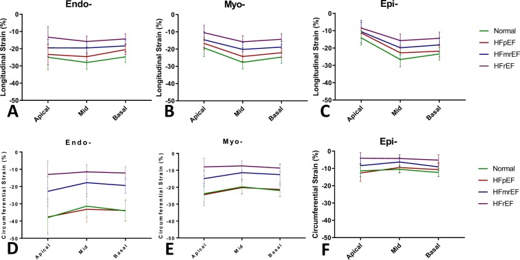
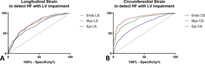
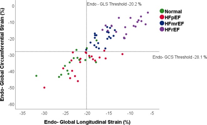
Heart failure (HF) is associated with progressive ventricular remodeling and impaired contraction that affects distinctly various regions of the myocardium. Our study applied cardiac magnetic resonance (CMR) feature tracking (FT) to assess comparatively myocardial strain at 3 distinct levels: subendocardial (Endo-), mid (Myo-) and subepicardial (Epi-) myocardium across an extended spectrum of patients with HF. 59 patients with HF, divided into 3 subgroups as follows: preserved ejection fraction (HFpEF, N = 18), HF with mid-range ejection fraction (HFmrEF, N = 21), HF with reduced ejection fraction (HFrEF, N = 20) and a group of age- gender- matched volunteers (N = 17) were included. Using CMR FT we assessed systolic longitudinal and circumferential strain and strain-rate at Endo-, Myo- and Epi- levels. Strain values were the highest in the Endo- layer and progressively lower in the Myo- and Epi- layers respectively, this gradient was present in all the patients groups analyzed but decreased progressively in HFmrEF and further on in HFrEF groups. GLS decreased with the severity of the disease in all 3 layers: Normal > HFpEF > HFmrEF > HFrEF (Endo-: -23.0 ± 3.5 > -20.0 ± 3.3 > -16.4 ± 2.2 > -11.0 ± 3.2, p < 0.001, Myo-: -20.7 ± 2.4 > -17.5.0 ± 2.6 > -14.5 ± 2.1 > -9.6 ± 2.7, p < 0.001; Epi-: -15.7 ± 1.9 > -12.2 ± 2.1 > -10.6 ± 2.3 > -7.7 ± 2.3, p < 0.001). In contrast, GCS was not different between the Normal and HFpEF (Endo-: -34.5 ± 6.2 vs -33.9 ± 5.7, p = 0.51; Myo-: -21.9 ± 3.8 vs -21.3 ± 2.2, p = 0.39, Epi-: -11.4 ± 2.0 vs -10.9 ± 2.3, p = 0.54) but was, as well, markedly lower in the systolic heart failure groups: Normal > HFmrEF > HFrEF (Endo-: -34.5 ± 6.2 > -20.0 ± 4.2 > 12.3 ± 4.2, p < 0.001; Myo-: -21.9 ± 3.8 > -13.0 ± 3.4 > -8.0 ± 2.7. p < 0.001; Epi-: -11.4 ± 2.0 > -7.9 ± 2.3 > -4.5 ± 1.9. p < 0.001). CMR feature tracking multilayer strain assessment identifies large range differences between distinct myocardial regions. Our data emphasizes the importance of sub-endocardial myocardium for cardiac contraction and thus, its predilect role in imaging detection of functional impairment. CMR feature tracking offers a convenient, readily available, platform to evaluate myocardial contraction with excellent spatial resolution, rendering further details about discrete areas of the myocardium. Using this technique across distinct groups of patients with heart failure (HF), we demonstrate that subendocardial regions of the myocardium exhibit much higher strain values than mid-myocardium or subepicardial and are more sensitive to detect contractile impairment. We also show comparatively higher values of circumferential strain compared with longitudinal and a higher sensitivity to detect contractile impairment. A newly characterized group of patients, HF with mid-range ejection fraction (EF), shows similar traits of decompensation but has relatively higher strain values as patients with HF with reduced EF.


Conflict of interest statement

The authors declare no competing interests.

Figures

Figure 1
Figure 1
Range Variability of Multilayer Myocardial Strain across Different Stages of Heart Failure. (A) CMR Feature Tracking Assessment of Multilayer Longitudinal Strain: multilayer Longitudinal Strain in representative subjects from the 4 pathology groups, from left to right: Normal, HFpEF, HFmrEF, HFrEF. For each case on the vertical, from up to down, are shown respectively:long- axis 4Ch Cine Image in end-diastole, Global Longitudinal Strain vs time curves at 3 distinct myocardial layers, underlined Endo-, Myo- and Epi- myocardial layers at which the strain values were assessed, long-axis 4Ch Cine Image in end-systole. (B) CMR Feature Tracking Assessment of Multilayer Circumferential Strain: multilayer Circumferential Strain in the same representative subjects from the 4 groups, from left to right: Normal, HFpEF, HFmrEF, HFrEF. For each case on the vertical, from up to down, are shown, respectively: short-axis Basal Cine Image in end-diastole, Global Circumferential Strain vs time curves at 3 distinct myocardial layers, underlined Endo-, Myo- and Epi- myocardial layers at which the strain values were assessed, short-axis Basal Cine Image in end-systole. (C) Global Longitudinal Srain and (D) Global Circumferential Strain across the 4 pathology groups: multilayer assessment of Endo-, Myo-, Epi- layers of left ventricular myocardium. Normal (formula image) normal age-, gender- matched control, HFpEF (formula image) patients with HF with preserved EF, HFmrEF (formula image) patients with HF with mid-range reduced EF, HFrEF (formula image) patients with HF with reduced EF.
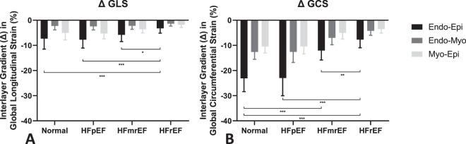
Figure 2
Figure 2
Interlayer Strain Gradient. Representation of (A) Global Longitudinal Strain and (B) Global Circumferential Strain Interlayer gradient (Δ) between Endo-Epi, Endo-Myo and Myo-Epi respectively in 4 groups: Normal – normal age-, gender- matched control, HFpEF – patients with HF with preserved EF, HFmrEF – patients with HF with mid-range reduced EF, HFrEF – patients with HF with reduced EF.
Figure 3
Figure 3
Regional Values of GLS and GCS across the 3 groups of patients with HF and Normal. (A) Endo-, (B) Myo- and (C) Epi- multilayer assessment of regional longitudinal strain and respectively (D) Endo-, (F) Myo- and (E) Epi- multilayer assessment of regional circumferential strain of left ventricular myocardium at 3 distinct ventricular levels Apical, Mid-ventricular and Basal in 4 groups: Normal – normal age-, gender- matched control, HFpEF – patients with HF with preserved EF, HFmrEF – patients with HF with mid-range EF, HFrEF – patients with HF with reduced EF.
Figure 4
Figure 4
Comparative ROC Analysis for Multilayer GLS and GCS Parameters. ROC analysis to discriminate patients with HF with LV impairment from Normal Subjects of multilayer. (A) Longitudinal Strain, and (B) Circumferential Strain (Endo LS – sub-endocardial longitudinal strain, Myo LS – mid-myocardial longitudinal strain, Epi LS, sub-epicardial longitudinal strain, Endo CS – sub-endocardial circumferential strain, Myo CS – mid-myocardial circumferential strain, Epi CS, sub-epicardial circumferential strain).
Figure 5
Figure 5
Distribution of HF Patients and Normal according to GLS and GCS Threshold Values. Scatterplot with Endo- Global Longitudinal Strain and Endo- Global Circumferential values. Reference lines represent the threshold values obtained through ROC analysis and Youden’s index calculation. The 4 groups represented are Normal – normal age-, gender- matched control, HFpEF – patients with HF with preserved EF, HFmrEF – patients with HF with mid-range reduced EF, HFrEF – patients with HF with reduced EF.

References

    1. Merlo M, et al. Long-term prognostic impact of therapeutic strategies in patients with idiopathic dilated cardiomyopathy: changing mortality over the last 30 years. Eur J Heart Fail. 2014;16:317–324. doi: 10.1002/ejhf.16. - DOI - PubMed
    1. Conrad N, et al. Temporal trends and patterns in heart failure incidence: a population-based study of 4 million individuals. Lancet. 2018;391:572–580. doi: 10.1016/S0140-6736(17)32520-5. - DOI - PMC - PubMed
    1. Writing Group M, et al. Heart Disease and Stroke Statistics-2016 Update: A Report From the American Heart Association. Circulation. 2016;133:e38–360. doi: 10.1161/CIR.0000000000000350. - DOI - PubMed
    1. Bogaert J, Rademakers FE. Regional nonuniformity of normal adult human left ventricle. Am J Physiol Heart Circ Physiol. 2001;280:H610–620. doi: 10.1152/ajpheart.2001.280.2.H610. - DOI - PubMed
    1. Buckberg G, Hoffman JI, Mahajan A, Saleh S, Coghlan C. Cardiac mechanics revisited: the relationship of cardiac architecture to ventricular function. Circulation. 2008;118:2571–2587. doi: 10.1161/CIRCULATIONAHA.107.754424. - DOI - PubMed

Publication types