. 2019 Nov 12:8:e47365.
doi: 10.7554/eLife.47365.

Interspecies interactions induce exploratory motility in Pseudomonas aeruginosa

Affiliations

Interspecies interactions induce exploratory motility in Pseudomonas aeruginosa

Dominique H Limoli et al. Elife. .

Abstract

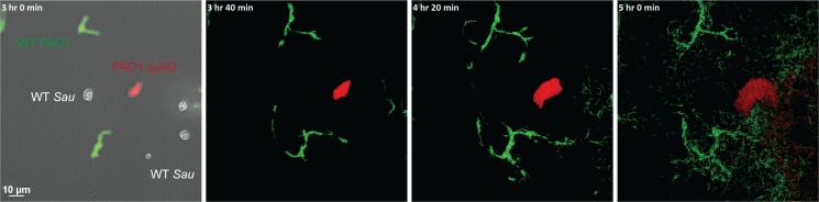
Microbes often live in multispecies communities where interactions among community members impact both the individual constituents and the surrounding environment. Here, we developed a system to visualize interspecies behaviors at initial encounters. By imaging two prevalent pathogens known to be coisolated from chronic illnesses, Pseudomonas aeruginosa and Staphylococcus aureus, we observed P. aeruginosa can modify surface motility in response to secreted factors from S. aureus. Upon sensing S. aureus, P. aeruginosa transitioned from collective to single-cell motility with an associated increase in speed and directedness - a behavior we refer to as 'exploratory motility'. Explorer cells moved preferentially towards S. aureus and invaded S. aureus colonies through the action of the type IV pili. These studies reveal previously undescribed motility behaviors and lend insight into how P. aeruginosa senses and responds to other species. Identifying strategies to harness these interactions may open avenues for new antimicrobial strategies.

Keywords: Pseudomonas aeruginosa; Staphylococcus aureus; bacterial motility; cystic fibrosis; infectious disease; microbiology; polymicrobial infections.


Conflict of interest statement

DL, EW, KY, ND, AC, GO No competing interests declared

Figures

Figure 1.
Figure 1.. S. aureus increases P. aeruginosa motility.
Live-imaging of polymicrobial interactions. P. aeruginosa (rod-shaped) was inoculated between a coverslip and an agarose pad, either in monoculture (top) or in coculture with equal numbers of S. aureus (cocci-shaped, bottom). Images were acquired every 15 m for 9 hr. Representative snap-shots of Video 1 (top) and Video 2 (bottom) are shown. Founding cells identified in the first frame are indicated with green rods (P. aeruginosa) or yellow circles (S. aureus). The location of the founding cell is indicated in each subsequent frame for positional reference. At t = 9 h:00 m, the founding P. aeruginosa cell has moved outside the field of view.
Figure 2.
Figure 2.. P. aeruginosa adopts an exploratory mode of motility in the presence of S. aureus.
Live-imaging of P. aeruginosa with WT S. aureus (Video 3). (A) Montage of representative snap-shots are shown beginning at 4 hr:28 m. Founding cells identified in the first frame are indicated with green rods (P. aeruginosa) or yellow circles (S. aureus). The location of the founding cell is indicated in each subsequent frame for positional reference. (B) Snap-shot at 5 hr, zoomed in to visualize single-cells. (C) Snap-shot at 6 hr of P. aeruginosa (mKO, red) and S. aureus (GFP, green) illustrating P. aeruginosa invasion into S. aureus colonies. (D) Snap-shot of coculture at 8 hr, showing disruption of S. aureus colonies (green arrows) and swift-moving P. aeruginosa cells out of the plane of focus (red arrows). Single P. aeruginosa cells and the leading edge of rafts were tracked in the presence of S. aureus and the speed (µm/s), acceleration (µm/s2), and mean squared displacement (µm2) for four independent videos are indicated, respectively, in (E – G).
Figure 3.
Figure 3.. P. aeruginosa exploratory motility is driven by type IV pili.
Live-imaging of P. aeruginosa with WT S. aureus. (A) Representative snap-shots of coculture with WT S. aureus and P. aeruginosa (WT, ΔpilA, ΔflgK, and ΔpilA ΔflgK, left to right, Videos 4 and 5 and Figure 3—videos 1 and 2 respectively) are shown at t = 4.5 hr. Boxed insets show swift-moving P. aeruginosa cells out of the plane of focus (red arrows). (B) The area of S. aureus per frame in monoculture or in the presence of the indicated P. aeruginosa strain was calculated at t = 5 hr by dividing the total area occupied by S. aureus in a single frame by the number of S. aureus colonies. A minimum of four videos were analyzed per condition. The mean and standard deviation are indicated. Statistical significance was determined by one-way ANOVA followed by Tukey’s Multiple Comparisons Test - a indicates a statistically significant difference (p≤0.05) between S. aureus in monoculture and in the presence of either WT P. aeruginosa or ΔflgK; b indicates a statistically significant difference between ΔpilA and ΔpilA ΔflgK. (C) Representative snap-shots of Video 4, WT P. aeruginosa and S. aureus beginning at 4 hr with 50 ms intervals, showing back-and-forth motion. Red arrows indicate when a P. aeruginosa cell is moving in towards the S. aureus colony and green arrows indicate when a cell is moving away.
Figure 3—figure supplement 1.
Figure 3—figure supplement 1.. Live-imaging of P. aeruginosa ΔpilA ΔflgK mutant in monoculture.
Representative snapshot at t = 4 hr.
Figure 4.
Figure 4.. Agr-regulated secreted S. aureus factors increase P. aeruginosa motility.
(A – D) Motility of WT P. aeruginosa was monitored by macroscopic sub-surface inoculation assays in the presence of medium alone or cell-free supernatant from the indicated S. aureus strains. (A) illustrates representative motility zones stained with crystal violet for visualization. The dilution factor of the supernatant is indicated in A and B. Undiluted supernatant was used in C. In D and E, the motility of the indicated P. aeruginosa mutants was analyzed in the presence of medium alone or supernatant derived from WT S. aureus (0.5 dilution) under 1.5% agar (D) or within 0.3% agar (E). The mean and standard deviation are indicated for at least three biological replicates. Statistical significance was determined by one-way ANOVA followed by Tukey’s Multiple Comparisons Test - a indicates a statistically significant difference (p≤0.05) between the motility observed in the presence of S. aureus supernatant compared to medium alone, and b indicates a statistically significant difference (p≤0.05) between the motility observed in the mutant strain (S. aureus mutants in B and C, P. aeruginosa mutants in D and E) compared to the parental.
Figure 5.
Figure 5.. P. aeruginosa biases the directionality of movement up a concentration gradient of Agr-regulated secreted factors.
(A) A concentration gradient of either S. aureus growth medium (TSB), S. aureus supernatant derived from WT or ΔagrBDCA was established by spotting onto the surface of the agar and allowing a concentration gradient to establish by diffusion for approximately 24 hr, prior to spotting P. aeruginosa onto the agar (6 mm to the left) and surface-based motility imaged after 24 hr. Representative images of at least three independent experiments are shown. (B) Example of live-imaging of WT P. aeruginosa with S. aureus ΔagrBDCA with tracks of single-cells shown and schematic illustrating the methods for calculating the directedness. Single P. aeruginosa cells were tracked from first the frame a cell exited the raft to the frame where it first encounters S. aureus. The accumulated track distance, D(A), was measured for at least 30 cells in four independent videos and compared to the Euclidean distance, D(E), between the position of the cell in the first and last frame tracked. The ratio of D(E)/D(A) (Directedness) is shown in (C) for P. aeruginosa moving towards WT S. aureus compared to ΔagrBDCA with the mean indicated. Statistical significance was determined by an unpaired Student’s t-test (a = P ≤ 0.05).
Figure 6.
Figure 6.. The CheY - like response regulator, PilG modulates P. aeruginosa response to S. aureus.
Representative snap-shots of P. aeruginosa ΔpilJ (A) and ΔpilG (B) in coculture with WT S. aureus. In (C), representative snap-shots of WT P. aeruginosa (GFP, green) with P. aeruginosa ΔpilG (mKate, red) and WT S. aureus (unmarked), visible only in the first frame (left) with phase contrast overlay.
Figure 6—figure supplement 1.
Figure 6—figure supplement 1.. Motility of WT, ΔpilA, ΔpilJ, and ΔpilG P. aeruginosa by macroscopic sub-surface inoculation assays in the presence of medium alone (TSB) or cell-free supernatant from WT S. aureus strains.
The mean and standard deviation are indicated for at least three biological replicates. Statistical significance was determined by one-way ANOVA followed by Tukey’s Multiple Comparisons Test - a indicates a statistically significant difference (p≤0.05) between the motility observed in the mutant P. aeruginosa strains compared to the parental (WT).
Figure 6—figure supplement 2.
Figure 6—figure supplement 2.. Live-imaging of PAO1 ΔpilG in coculture with WT PAO1 and WT S. aureus.
Representative snap-shots of P. aeruginosa WT PAO1 (GFP, green), PAO1 ΔpilG (mKate, red) in coculture with WT S. aureus (phase contrast only, in first frame, left) showing diminished response to S. aureus in comparison to WT PAO1.
Figure 6—figure supplement 3.
Figure 6—figure supplement 3.. Live-imaging of PA14 ΔpilG in coculture with increased S. aureus inoculum.
Representative snap-shots of P. aeruginosa ΔpilG in coculture with WT S. aureus showing increased S. aureus inoculum promotes motility in a ΔpilG mutant.
Figure 7.
Figure 7.. P. aeruginosa modulates motility in response to a range of CF clinical and non-clinical bacterial species.
The motility of clinical P. aeruginosa isolates (CFBR PA 38, 43, 44, and 37), in the presence of cell-free supernatant derived from S. aureus USA300 LAC (0.5 dilution) is indicated, in comparison to P. aeruginosa PA14 (A). The motility of P. aeruginosa laboratory strain PA14 was monitored in the presence of undiluted cell-free supernatant from clinical S. aureus CF isolates, CFBR SA 47, 48, and 50 (B), CF clinical isolates: H. influenzae, B. cepacia, and A. xylosoxidans (C), and non-CF species: E. coli, B. subtilis, and S. Typhimurium (D). Growth medium alone for each species was used as a negative control (TSB: S. aureus, BHI + Fildes: H. influenzae, LB: B. cepacia, A. xylosoxidans, B. subtilis, E. coli, and S. Typhimurium). The mean and standard deviation are indicated for at least three biological replicates. Statistical significance was determined by one-way ANOVA followed by Tukey’s Multiple Comparisons Test – a indicates a statistically significant difference (p≤0.05) between the motility observed in the presence of S. aureus supernatant compared to medium alone (A–D), and b indicates a statistically significant difference (p≤0.05) between the motility observed in the CF isolates, in comparison to laboratory strains (P. aeruginosa laboratory strain PA14 in (A) and S. aureus USA300 LAC in (B)).
Figure 7—figure supplement 1.
Figure 7—figure supplement 1.. Clinical CF S. aureus isolates retain β-hemolysis.
Laboratory S. aureus isolates USA300 LAC (WT) and ΔagrBDCA and clinical CF S. aureus isolates (CFBRSA47, 48, and 50) were grown on sheep’s blood agar and examined for hemolysis of red blood cells surrounding the colonies.
Figure 8.
Figure 8.. Phenol soluble modulins contribute to S. aureus-induced P. aeruginosa motility.
Motility of WT P. aeruginosa was monitored by macroscopic sub-surface inoculation assays in the presence of medium alone or cell-free supernatant derived from WT S. aureus treated with either heat, proteinase K (PK), or trypsin (tryp) in (A), methanol/chloroform extraction of the supernatant and subsequent analysis of the organic and aqueous fraction (methanol-chloroform (2:1, Me/Chlor) was included as the vehicle control) (B), or in the presence of untreated supernatant derived from WT or the indicated S. aureus mutants (0.5 dilution) in (C). The mean and standard deviation are indicated for at least three biological replicates. Statistical significance was determined by one-way ANOVA followed by Tukey’s Multiple Comparisons Test - a indicates a statistically significant difference (p≤0.05) between the motility observed in the presence of S. aureus supernatant compared to medium alone, b between the motility observed in the mutant strains compared to the parental, and c between the ΔagrBDCA and Δpsmαβδ mutants.

References

    1. Anyan ME, Amiri A, Harvey CW, Tierra G, Morales-Soto N, Driscoll CM, Alber MS, Shrout JD. Type IV pili interactions promote intercellular association and moderate swarming of Pseudomonas aeruginosa. PNAS. 2014;111:18013–18018. doi: 10.1073/pnas.1414661111. - DOI - PMC - PubMed
    1. Arnaud M, Chastanet A, Débarbouillé M. New vector for efficient allelic replacement in naturally nontransformable, low-GC-content, Gram-positive bacteria. Applied and Environmental Microbiology. 2004;70:6887–6891. doi: 10.1128/AEM.70.11.6887-6891.2004. - DOI - PMC - PubMed
    1. Bæk KT, Frees D, Renzoni A, Barras C, Rodriguez N, Manzano C, Kelley WL. Genetic variation in the Staphylococcus aureus 8325 strain lineage revealed by whole-genome sequencing. PLOS ONE. 2013;8:e77122. doi: 10.1371/journal.pone.0077122. - DOI - PMC - PubMed
    1. Bertrand JJ, West JT, Engel JN. Genetic analysis of the regulation of type IV pilus function by the Chp chemosensory system of Pseudomonas aeruginosa. Journal of Bacteriology. 2010;192:994–1010. doi: 10.1128/JB.01390-09. - DOI - PMC - PubMed
    1. Bligh EG, Dyer WJ. A rapid method of total lipid extraction and purification. Canadian Journal of Biochemistry and Physiology. 1959;37:911–917. doi: 10.1139/y59-099. - DOI - PubMed

Publication types

MeSH terms

LinkOut - more resources