

Working memory training and brain structure and function in extremely preterm or extremely low birth weight children
- PMID: 31713952
- PMCID: PMC6977425
- DOI: 10.1002/hbm.24832
Working memory training and brain structure and function in extremely preterm or extremely low birth weight children
Abstract
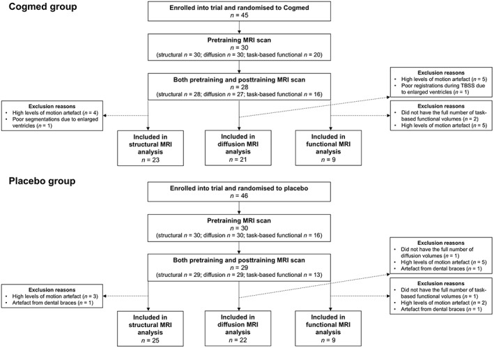
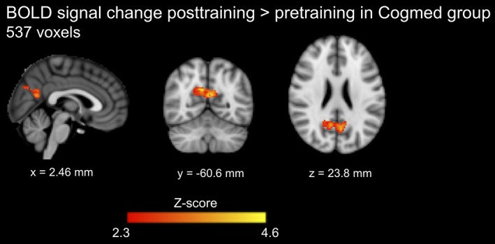
This study in children born extremely preterm (EP; <28 weeks' gestational age) or extremely low birth weight (ELBW; <1,000 g) investigated whether adaptive working memory training using Cogmed® is associated with structural and/or functional brain changes compared with a placebo program. Ninety-one EP/ELBW children were recruited at a mean (standard deviation) age of 7.8 (0.4) years. Children were randomly allocated to Cogmed or placebo (45-min sessions, 5 days a week over 5-7 weeks). A subset had usable magnetic resonance imaging (MRI) data pretraining and 2 weeks posttraining (structural, n = 48; diffusion, n = 43; task-based functional, n = 18). Statistical analyses examined whether cortical morphometry, white matter microstructure and blood oxygenation level-dependent (BOLD) signal during an n-back working memory task changed from pretraining to posttraining in the Cogmed and placebo groups separately. Interaction analyses between time point and group were then performed. There was a significant increase in neurite density in several white matter regions from pretraining to posttraining in both the Cogmed and placebo groups. BOLD signal in the posterior cingulate and precuneus cortices during the n-back task increased from pretraining to posttraining in the Cogmed but not placebo group. Evidence for group-by-time interactions for the MRI measures was weak, suggesting that brain changes generally did not differ between Cogmed and placebo groups. Overall, while some structural and functional MRI changes between the pretraining and posttraining period in EP/ELBW children were observed, there was little evidence of training-induced neuroplasticity, with changes generally identified in both groups. Trial registration Australian New Zealand Clinical Trials Registry, anzctr.org.au; ACTRN12612000124831.
Keywords: Cogmed; diffusion imaging; functional imaging; magnetic resonance imaging; microstructure; preterm birth.
© 2019 The Authors. Human Brain Mapping published by Wiley Periodicals, Inc.
Conflict of interest statement
The authors declare no conflicts of interest.
Figures


References
-
- Anderson, P. J. (2014). Neuropsychological outcomes of children born very preterm. Seminars in Fetal & Neonatal Medicine, 19, 90–96. - PubMed
-
- Anderson, P. J. , Doyle, L. W. , & Victorian Infant Collaborative Study Group . (2004). Executive functioning in school‐aged children who were born very preterm or with extremely low birth weight in the 1990s. Pediatrics, 114, 50–57. - PubMed
-
- Anderson, P. J. , Lee, K. J. , Roberts, G. , Spencer‐Smith, M. M. , Thompson, D. K. , Seal, M. L. , … Pascoe, L. (2018). Long‐term academic functioning following Cogmed working memory training for children born extremely preterm: A randomized controlled trial. The Journal of Pediatrics, 202, 92–97. - PubMed
-
- Andersson, J. L. , Skare, S. , & Ashburner, J. (2003). How to correct susceptibility distortions in spin‐echo echo‐planar images: Application to diffusion tensor imaging. NeuroImage, 20, 870–888. - PubMed
