

Peak Sinus Pressures During Sneezing in Healthy Controls and Post-Skull Base Surgery Patients
- PMID: 31714627
- PMCID: PMC7549275
- DOI: 10.1002/lary.28400
Peak Sinus Pressures During Sneezing in Healthy Controls and Post-Skull Base Surgery Patients
Abstract
Objectives/hypothesis: Patients are frequently advised to sneeze with an open mouth and avoid nose-blowing following an endoscopic endonasal approache (EEA) to the skull base, despite a lack of quantitative evidence. This study applies computational fluid dynamics (CFD) to quantify sinus pressures along the skull base during sneezing.
Study design: Case-control series.
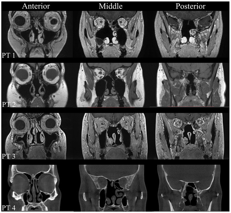
Methods: Computed tomography or magnetic resonance imaging scans of four post-EEA patients and four healthy controls were collected and analyzed utilizing CFD techniques. A pressure drop of 6,000 Pa was applied to the nasopharynx based on values in the literature to simulate expiratory nasal airflow during sneezing. Peak pressures along the skull base in frontal, ethmoid, and sphenoid sinuses were collected.
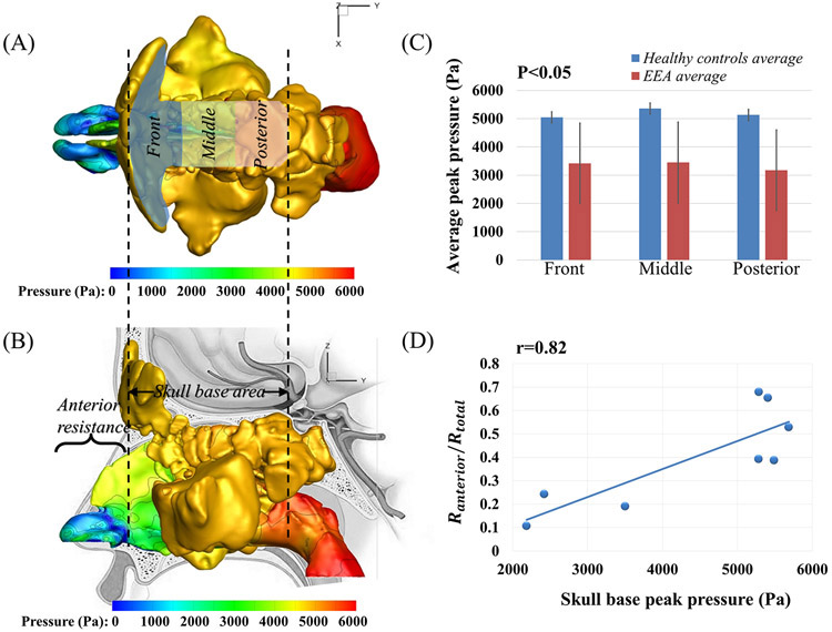
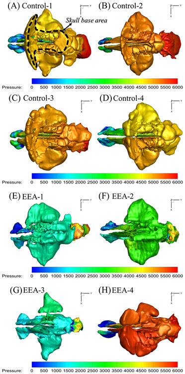
Results: Significant increases in skull base peak pressure was observed during sneezing, with significant individual variations from 2,185 to 5,685 Pa. Interestingly, healthy controls had significantly higher pressures compared to post-EEA patients (5179.37 ± 198.42 Pa vs. patients 3,347.82 ± 1,472.20 Pa, P < .05), which could be related to higher anterior nasal resistance in unoperated healthy controls (0.44 ± 0.22 vs. 0.31 ± 0.16 Pa/mL/sec for patients, P = .38). The sinus pressure buildup may be due to airway resistance functioning as a valve preventing air from being released quickly. Supporting this theory, there was a strong correlation (r = 0.82) between peak skull base pressure and the ratio of anterior resistance to total resistance. Within-subject variation in pressures between different skull base regions was much lower (average = ~5%).
Conclusions: This study provided the first quantitative analysis of air pressure along the skull base during sneezing in post-EEA patients through CFD, suggesting that pressure buildup may depend on individual anatomy.
Level of evidence: 3b Laryngoscope, 130:2138-2143, 2020.
Keywords: Skull base surgery; computational fluid dynamics; nasal airflow dynamics.
© 2019 The American Laryngological, Rhinological and Otological Society, Inc.
Conflict of interest statement
The authors have no financial interest and conflict of interest to disclose.
Figures
References
-
- Cappabianca P, Cavallo LM, Esposito F, de Divitiis O, Messina A, de Divitiis E. Extended endoscopic endonasal approach to the midline skull base: the evolving role of transsphenoidal surgery In: Pickard JD, Akalan N, Di Rocco C, et al., eds. Advances and Technical Standards in Neurosurgery. Vienna: Springer Vienna; 2008:151–199. doi:10.1007/978-3-211-72283-1_4 - DOI - PubMed
Publication types
MeSH terms
Grants and funding
LinkOut - more resources
Full Text Sources
Medical
Research Materials
Miscellaneous
