. 2019 Nov 12;14(11):e0223254.
doi: 10.1371/journal.pone.0223254. eCollection 2019.

SNARE proteins rescue impaired autophagic flux in Down syndrome

Affiliations

SNARE proteins rescue impaired autophagic flux in Down syndrome

Stefanos Aivazidis et al. PLoS One. .

Abstract

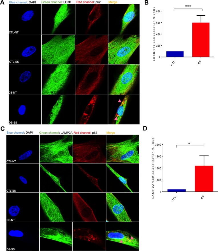
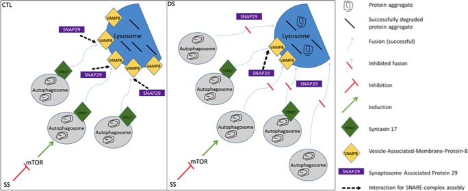
Down syndrome (DS) is a chromosomal disorder caused by trisomy of chromosome 21 (Ts21). Unbalanced karyotypes can lead to dysfunction of the proteostasis network (PN) and disrupted proteostasis is mechanistically associated with multiple DS comorbidities. Autophagy is a critical component of the PN that has not previously been investigated in DS. Based on our previous observations of PN disruption in DS, we investigated possible dysfunction of the autophagic machinery in human DS fibroblasts and other DS cell models. Following induction of autophagy by serum starvation, DS fibroblasts displayed impaired autophagic flux indicated by autophagolysosome accumulation and elevated p62, NBR1, and LC3-II abundance, compared to age- and sex-matched, euploid (CTL) fibroblasts. While lysosomal physiology was unaffected in both groups after serum starvation, we observed decreased basal abundance of the Soluble N-ethylmaleimide-sensitive-factor Attachment protein Receptor (SNARE) family members syntaxin 17 (STX17) and Vesicle Associated Membrane Protein 8 (VAMP8) indicating that decreased autophagic flux in DS is due at least in part to a possible impairment of autophagosome-lysosome fusion. This conclusion was further supported by the observation that over-expression of either STX17 or VAMP8 in DS fibroblasts restored autophagic degradation and reversed p62 accumulation. Collectively, our results indicate that impaired autophagic clearance is a characteristic of DS cells that can be reversed by enhancement of SNARE protein expression and provides further evidence that PN disruption represents a candidate mechanism for multiple aspects of pathogenesis in DS and a possible future target for therapeutic intervention.


Conflict of interest statement

The authors have declared that no competing interests exist.

Figures

Fig 1
Fig 1. The proteostasis network in Down syndrome.
The pathways responsible for preserving proteostasis constitute the proteostasis network (PN). The PN includes the ribosome for proper protein translation, molecular chaperones (HSP) for correct protein folding and protein degradation machinery (26S proteasome for individual peptide degradation and ER-associated degradation (ERAD), autophagy for bulk protein degradation). Its major processes involve successful synthesis, folding, repair/disaggregation and degradation of proteins. In addition, there are several secondary PN modulators, like the unfolded protein response (UPR) caused by endoplasmic reticulum (ER) stress that constitute the stress responsive arm of the PN and provide its plasticity. For extensive reviews of the PN see the following references [, –85].
Fig 2
Fig 2. DS fibroblasts exhibit increased autophagolysosome accumulation and elevated abundance of p62 and LC3II after serum starvation.
(A) Representative blot of LC3-I, LC3-II, p62 and β-actin of a CTL (AG004392) and DS fibroblast (AG006872) cell line (three technical replicates per treatment and genotype are presented for each cell line). (B) Quantification of abundance levels of LC3-II and p62 at basal levels (% based on abundance of CTL NT) (C) Quantification of fold change in abundance of LC3II and p62 after serum starvation. (D) Immunofluorescence for p62 (Red) and DAPI (blue) following serum starvation of CTL and DS fibroblasts. (E) Quantification of p62 fluorescence intensity (% based on abundance of CTL serum starvation). (F) TEM figures of CTL and DS fibroblasts following serum starvation. (G) Quantitation of autophagolysosome number in the TEM figures. AL, autophagolysosome; AP, autophagosome; M, mitochondria; SS, serum starvation (8h).
Fig 3
Fig 3. Colocalization of p62 with LC3B and LAMP2A confirms the presence of undegraded autophagosomes and autophagolysosomes.
(A) IF for LC3B (Green), p62 (Red) and DAPI (Blue) in a CTL (AG004392) and DS (AG006872) fibroblast cell line at basal levels or after serum starvation. (B) Quantification of LC3B/p62 co-localization (yellow) fluorescence intensity (area-pixels) after serum starvation (% based on abundance of CTL serum starvation). Pink arrow = p62/LC3B co-localized puncta. NT, Not treated-basal levels; SS, serum starvation (8h). (C) IF for LAMP2A (Green), p62 (Red) and DAPI (Blue) in a CTL and DS fibroblast cell line at basal levels or after serum starvation. (D) Quantification of LAMP2A/p62 co-localization (yellow) fluorescence intensity (area-pixels) after serum starvation (% based on abundance of CTL serum starvation). Pink arrow = p62/LAMP2A co-localized puncta.
Fig 4
Fig 4. Lysosomal dysfunction is not responsible for p62 accumulation in DS fibroblasts after serum starvation.
(A) Representative blot of LC3-I, LC3-II, p62 and β-actin of a CTL (AG004392) and DS (AG006872) fibroblast cell line at basal levels, after CQ treatment (dosage: 30 um), after serum starvation and CQ co-treatment and after serum starvation and H2O co-treatment used as control. (B) Quantification of fold change in abundance of p62 of a CTL and DS fibroblast cell line after CQ, serum starvation+CQ and serum starvation+H2O. (C) Quantification of fold change in abundance of LC3-II of a CTL and DS fibroblast cell line after CQ, serum starvation+CQ and serum starvation+H2O. (D) Lysotracker-Red fluorescence intensity in a CTL and DS fibroblast cell line after serum starvation (% based on fluorescence intensity of CTL serum starvation). (E) Proteolytic activity of lysosomal cathepsin B (% based on activity of CTL serum starvation).
Fig 5
Fig 5. Overexpression of STX17 and VAMP8 restores autophagic flux in DS cells after serum starvation.
(A) Representative blot of STX17 and β-actin or VAMP8 and β-actin of CTL and DS fibroblast cell line at basal levels (NT) or after serum starvation (2h-8h). (B) Quantification of STX17 and VAMP8 abundance (%CTL) at basal levels. (C) IF in a CTL (AG004392) and DS (AG006872) fibroblast cell line CTL and DS fibroblast cell line after serum starvation for FLAG-only, FLAG-STX17, EGFP-only, EGFP-VAMP8 (green), p62 (Red) and DAPI (Blue). (D) Quantification of p62 fluorescence intensity (area-pixels) after serum starvation in DS (AG006872) fibroblast cell line transfected with EGFP-VAMP8 vector (% based on p62 abundance of CTL cells transfected with EGFP-only vector). Quantification of p62 fluorescence intensity (area-pixels) after serum starvation and in a DS (AG006872) fibroblast cell line transfected with FLAG-STX17 vector (% based on abundance of CTL cells transfected with FLAG-only EV) (E) Quantification of p62 fluorescence intensity (area-pixels) after serum starvation in DS (AG006872) fibroblasts transfected with FLAG-only or FLAG-STX17 vector (% based on abundance of DS cells transfected with FLAG-only vector). Quantification of p62 fluorescence intensity (area-pixels) after serum starvation in DS (AG006872) fibroblasts transfected with EGFP-only or EGFP-VAMP8 vector (% based on abundance of DS cells transfected with FLAG-only vector).
Fig 6
Fig 6. Diminished autophagic flux in DS fibroblasts after serum starvation is characterized by autophagosome fusion defects due to reduced STX17 and VAMP8 levels (SS = serum starvation, 8h).

References

    1. Wiseman FK, Al-Janabi T, Hardy J, Karmiloff-Smith A, Nizetic D, Tybulewicz VL, et al. A genetic cause of Alzheimer disease: mechanistic insights from Down syndrome. Nat Rev Neurosci. 2015;16(9):564–74. 10.1038/nrn3983 - DOI - PMC - PubMed
    1. Boghossian NS, Hansen NI, Bell EF, Stoll BJ, Murray JC, Carey JC, et al. Mortality and morbidity of VLBW infants with trisomy 13 or trisomy 18. Pediatrics. 2014;133(2):226–35. Epub 2014/01/20. 10.1542/peds.2013-1702 - DOI - PMC - PubMed
    1. Wang X, Huang T, Bu G, Xu H. Dysregulation of protein trafficking in neurodegeneration. Molecular neurodegeneration. 2014;9:31 10.1186/1750-1326-9-31 - DOI - PMC - PubMed
    1. Blom NA, Ottenkamp J, Wenink AG, Gittenberger-de Groot AC. Deficiency of the vestibular spine in atrioventricular septal defects in human fetuses with down syndrome. The American journal of cardiology. 2003;91(2):180–4. 10.1016/s0002-9149(02)03106-5 . - DOI - PubMed
    1. Aitken RJ, Mehers KL, Williams AJ, Brown J, Bingley PJ, Holl RW, et al. Early-onset, coexisting autoimmunity and decreased HLA-mediated susceptibility are the characteristics of diabetes in Down syndrome. Diabetes Care. 2013;36(5):1181–5. 10.2337/dc12-1712 - DOI - PMC - PubMed

Publication types