. 2019 Nov 12;14(11):e0224621.
doi: 10.1371/journal.pone.0224621. eCollection 2019.

Identification of a novel monocytic phenotype in Classic Hodgkin Lymphoma tumor microenvironment

Affiliations

Identification of a novel monocytic phenotype in Classic Hodgkin Lymphoma tumor microenvironment

Ginell R Post et al. PLoS One. .

Abstract

Classic Hodgkin lymphoma (CHL) characteristically shows few malignant cells in a microenvironment comprised of mixed inflammatory cells. Although CHL is associated with a high cure rate, recent studies have associated poor prognosis with absolute monocyte count in peripheral blood and increased monocyte/macrophages in involved lymph nodes. Thus, the role of monocytic infiltration and macrophage differentiation in the tumor microenvironment of CHL may be more relevant than absolute macrophage numbers to defining prognosis in CHL patients and potentially have therapeutic implications. Most studies identify tumor-associated macrophages (TAMs) using markers (e.g., CD68) expressed by macrophages and other mononuclear phagocytes, such as monocytes. In contrast, Class A Scavenger Receptor (SR-A/CD204) is expressed by tissue macrophages but not monocytic precursors. In this study, we examined SR-A expression in CHL (n = 43), and compared its expression with that of other macrophage markers. We confirmed a high prevalence of mononuclear cells that stained with CD68, CD163, and CD14 in CHL lymph nodes. However, SR-A protein expression determined by immunohistochemistry was limited to macrophages localized in sclerotic bands characteristic of nodular sclerosis CHL. In contrast, SR-A protein was readily detectable in lymph nodes with metastatic tumor, extra-nodal CHL, T cell/histiocyte-rich large B cell lymphoma, and resident macrophages in non-malignant tissues, including spleen, lymph node, liver and lung. The results of SR-A protein expression paralleled the expression of SR-A mRNA determined by quantitative RT-PCR. These data provide evidence that tumor-infiltrating monocyte/macrophages in CHL have a unique phenotype that likely depends on the microenvironment of nodal CHL.


Conflict of interest statement

The authors have declared that no competing interests exist.

Figures
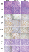
Fig 1
Fig 1. Expression pattern of monocyte/macrophage markers in nodal CHL.
Shown are hematoxylin and eosin (H&E) and immunostaining patterns of different cellular markers in FFPE sections from representative CHL morphologic variants [nodular sclerosis (NSCHL, left panels), mixed cellularity (MCCHL, middle panels) and lymphocyte rich (LRCHL, right panels)]. CD30 immunoreactivity was used to identify malignant HRS cells. Antibodies to CD68, CD163 and CD14 were used to stain monocyte/macrophages, and staining patterns for these antibodies compared with that of SR-A using two different monoclonal antibodies (clones OTI9ES and UMAB246). Notably, monocyte/macrophages in proximity to the malignant cells in nodal CHL were positive for these three monocyte/macrophage markers. In contrast, antibody staining for SR-A in nodal CHL was limited to the fibrotic bands characteristic of NSCHL.
Fig 2
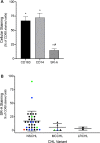
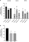
Fig 2. Analysis of CD163, CD14, and SR-A expression compared to CD68 in CHL lymph nodes.
Tissue sections of nodal CHL stained with CD68, CD163, CD14, and SR-A (OTI9ES) were scored by a pathologist. (A) The graph depicts the percent of CD68 positive cells that were also stained with CD163, CD14, or SR-A. * denotes significant differences (p<0.05) from CD68 assessed by one-way t-test; # denotes significant difference (p<0.05) from CD163 and CD14 determined by one-way ANOVA and Tukey’s post-hoc test. (B) SR-A staining relative to CD68 in nodal CHL based on morphologic subtype. As shown in Fig 1, SR-A staining of the NSCHL was detected in fibrotic bands (Green symbol: EBV positive cases; Blue symbol: relapsed disease; Red symbol: EBV positive, relapsed disease).
Fig 3
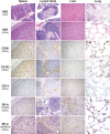
Fig 3. Expression pattern of monocyte/macrophage markers in spleen, lymph node, liver, and lung.
Shown are representative H&E and immunostaining patterns of resident tissue monocyte/macrophages in FFPE sections of spleen, lymph node, liver, and lung. SR-A staining (OTI9ES and UMAB246) shows a similar pattern to CD68 and CD163 in the splenic cords and sinus histiocytes (lymph nodes), Kuppfer cells (liver) and alveolar macrophages (lung). CD14 staining was different from the other macrophages markers in splenic red pulp cords, Kuppfer cells (liver), and alveolar macrophages (lung).
Fig 4
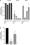
Fig 4. Analysis of CD163, CD14 and SR-A expression compared to CD68 in resident tissue monocyte/macrophages.
A) Tissue-resident monocyte/macrophages in FFPE sections of spleen, lymph node, liver, and lung were immunostained and scored for the percent of CD68 positive cells that also stained with CD163, CD14, or SR-A (OTI9ES). Symbols represent the percent of CD68 positive cells that were co-stained in individual samples. Bars depict the means and range for the individual tissue type. B) Average of CD68 positive monocyte/macrophages that also stained with CD163, CD14, or SR-A across tissue types. Bars depict the mean and SEM. * denotes significant differences (p<0.05) from CD68 assessed by one-way t-test; # denotes significant difference (p<0.05) from CD163 determined by one-way ANOVA and Tukey’s post-hoc test.
Fig 5
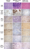
Fig 5. Expression pattern of monocyte/macrophage markers in nodal T cell/histiocyte-rich large B cell Lymphoma (THRLBCL), nodal metastatic colon cancer, and hepatic CHL.
Shown are representative H&E and immunostaining patterns of monocyte/macrophages in FFPE sections of nodal THRLBCL (left panels), nodal metastatic colon carcinoma (center panels) and extra-nodal CHL (right panels). CD20 identifies malignant B cells in THRLBCL, cytokeratin (CK) shows malignant metastatic tumor cells, and CD30 detects malignant HRS cells in CHL. SR-A staining (OTI9ES and UMAB246) shows a similar pattern to CD68, CD163 and CD14 in each tissue.
Fig 6
Fig 6. Analysis of CD163, CD14, and SR-A expression in TAMs.
A) Tumor-associated macrophages present in sections of THRLBCL, metastatic colon cancer, and hepatic CHL were immunostained and scored for the percent of CD68 positive cells that also stained with CD163, CD14, or SR-A (OTI9ES). Symbols represent the percent of CD68 positive cells that were co-stained in individual samples. Bars depict the means and range for the individual tumor types. B) Average of CD68 positive TAMs that also stained with CD163, CD14, or SR-A across tumor types. Bars depict the mean and SEM. * denotes significant differences (p<0.05) from CD68 assessed by one-way t-test; # denotes significant difference (p<0.05) from CD163 determined by one-way ANOVA and Tukey’s post-hoc test.
Fig 7
Fig 7. CD163, CD14 and SR-A expression in nodal CHL, lung and liver assessed by qRT-PCR.
Total RNA was prepared from FFPE sections of nodal NSCHL (n = 2) and MCCHL (n = 2), from tissue (lung and liver), and differentiated THP-1 macrophages (positive control). After reverse transcription, qRT-PCR was used to quantify expression of CD68, CD163, and SR-A. Relative expression of CD163 and SR-A in macrophages was calculated by normalizing their Ct values to that of CD68 in the same sample (ΔCt), and then to their expression in THP-1 macrophages (ΔΔCt). CD163 expression was readily detected in nodal CHL and lung/liver (tissue), and to a lesser extent in NSCHL and MCCHL. SR-A expression was readily detected in tissue, but was only detected at very low levels in NSCHL and not detected (ND) in MCCHL.

Similar articles

Cited by

References

    1. Kuppers R. The biology of Hodgkin's lymphoma. Nat Rev Cancer. 2009;9(1):15–27. 10.1038/nrc2542 . - DOI - PubMed
    1. Aldinucci D, Gloghini A, Pinto A, De Filippi R, Carbone A. The classical Hodgkin's lymphoma microenvironment and its role in promoting tumour growth and immune escape. The Journal of pathology. 2010;221(3):248–63. 10.1002/path.2711 . - DOI - PubMed
    1. Guo B, Cen H, Tan X, Ke Q. Meta-analysis of the prognostic and clinical value of tumor-associated macrophages in adult classical Hodgkin lymphoma. BMC Med. 2016;14(1):159 10.1186/s12916-016-0711-6 . - DOI - PMC - PubMed
    1. Steidl C, Lee T, Shah SP, Farinha P, Han G, Nayar T, et al. Tumor-associated macrophages and survival in classic Hodgkin's lymphoma. N Engl J Med. 2010;362(10):875–85. 10.1056/NEJMoa0905680 - DOI - PMC - PubMed
    1. Koh YW, Park C, Yoon DH, Suh C, Huh J. CSF-1R expression in tumor-associated macrophages is associated with worse prognosis in classical Hodgkin lymphoma. Am J Clin Pathol. 2014;141(4):573–83. Epub 2014/03/13. 10.1309/AJCPR92TDDFARISU . - DOI - PubMed

Publication types