. 2019 Nov 8;8(11):1406.
doi: 10.3390/cells8111406.

Dyskerin Mutations Present in Dyskeratosis Congenita Patients Increase Oxidative Stress and DNA Damage Signalling in Dictyostelium Discoideum

Affiliations

Dyskerin Mutations Present in Dyskeratosis Congenita Patients Increase Oxidative Stress and DNA Damage Signalling in Dictyostelium Discoideum

Javier Rodriguez-Centeno et al. Cells. .

Abstract

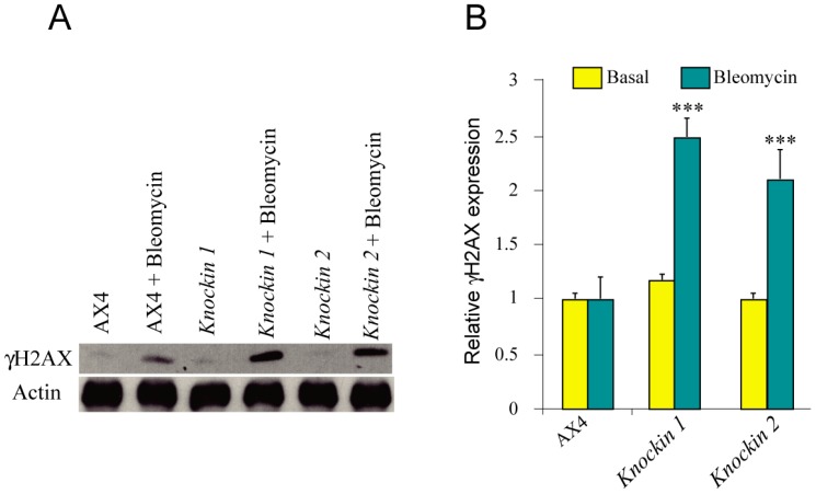
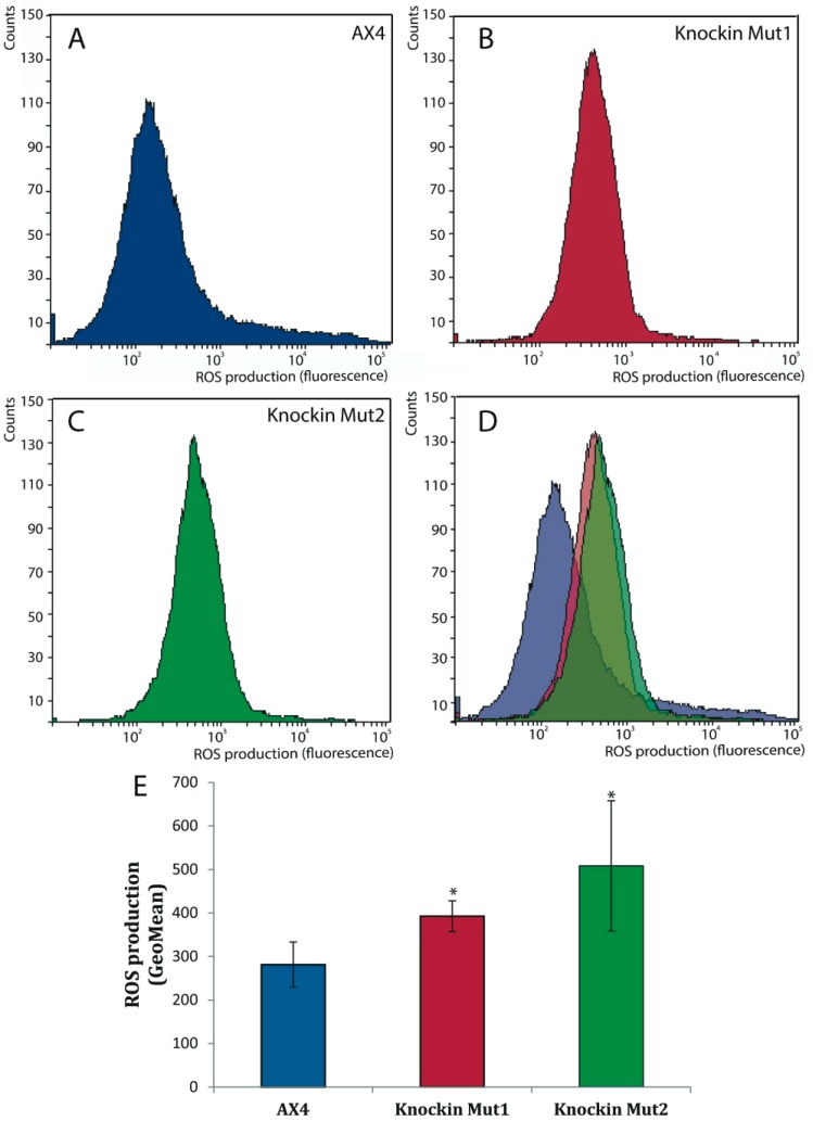
Dyskerin is a protein involved in the formation of small nucleolar and small Cajal body ribonucleoproteins. These complexes participate in RNA pseudouridylation and are also components of the telomerase complex required for telomere elongation. Dyskerin mutations cause a rare disease, X-linked dyskeratosis congenita, with no curative treatment. The social amoeba Dictyostelium discoideum contains a gene coding for a dyskerin homologous protein. In this article D. discoideum mutant strains that have mutations corresponding to mutations found in dyskeratosis congenita patients are described. The phenotype of the mutant strains has been studied and no alterations were observed in pseudouridylation activity and telomere structure. Mutant strains showed increased proliferation on liquid culture but reduced growth feeding on bacteria. The results obtained indicated the existence of increased DNA damage response and reactive oxygen species, as also reported in human Dyskeratosis congenita cells and some other disease models. These data, together with the haploid character of D. discoideum vegetative cells, that resemble the genomic structure of the human dyskerin gene, located in the X chromosome, support the conclusion that D. discoideum can be a good model system for the study of this disease.

Keywords: DNA damage; dictyostelium; dyskeratosis congenita; dyskerin; oxidative stress; pseudouridylation; telomere; telomere biology disorder.


Conflict of interest statement

The authors declare no conflict of interest.

Figures

Figure A1
Figure A1
Dkc1 expression in clones obtained by homologous recombination. RNA was isolated from different clones that had incorporated dkc1 by homologous recombination, as determined by the PCR test shown in Figure 2. The relative expression of dkc1 mRNA was determined by reverse transcription and quantitative PCR and compared to that of AX4 cells. Panel A shows the results obtained for several clones corresponding to the knockin-1 generation. Clone numbers are shown in the X-axes. The arrow indicates the clone that was selected for further analysis. Panel B shows the relative expression of the knockin-1 and knockin-2 clones selected for the study after removal of the Blasticidin resistance cassette upon expression of the Cre recombinase. This result is in agreement with the data on dkc1 expression shown in Figure 6.
Figure 1
Figure 1
Protein sequence and expression of D. discoideum dyskerin, (A) Schematic representation of dyskerin conserved domains. The conserved domains N-terminal extension (NTE), dyskerin-like domain (DSKLD), pseudouridin synthase catalytic domain (TruB_N) and RNA-binding domain (PUA) are represented according to the human protein. Amino acid positions are shown in the upper part of the diagram. (B) Comparison of the human (Hs) and D. discoideum (Dd) dyskerin amino acid sequences. Functional conserved domains are indicated using the same color code as in Panel A. The amino acids mutated in the D. discodeum protein in this study are highlighted in yellow. (C) D. discoideum nucleoli, one or two in each cell, were identified in the left panel by hybridization with a probe specific for the 26S ribosomal RNA. The right panel shows the expression of a D. discoideum dyskerin-GFP fusion protein after transfection of the cells with a dyskerin expression vector. Nuclei were stained with DAPI and are shown in blue color. Scale bars correspond to a distance of 5 μm.
Figure 2
Figure 2
Generation of dck1 knockin strains, (A) Schematic representation of the strategy used for the generation of dkc1 knockin strains by homologous recombination. The genomic organization of the dkc1 locus is represented to scale in the upper part indicating the dkc1 gene, the upstream DDB0308306 gene and the intergenic region that contains the putative dkc1 promoter region. The vector used for homologous recombination is represented underneath and includes the blasticidin resistance cassette (BSR) flanked by two Lox sites. Mutated residues are represented as red asterisks on the dkc1 gene. The product of homologous recombination is represented bellow. The location of the oligonucleotides used for verification of homologous recombination is indicated. The Bsr oligonucleotide hybridizes into the blasticidin-resistance cassette while the DKCK5 oligonucleotide correspond to a region of the BBB0308306 genes located upstream of the 5′ arm of the recombination vector. The position of the oligonucleotides used for amplification and sequencing of the mutated region, SeqF and SeqR, is also indicated. The lowest scheme represents the structure of the locus after removal of the BSR cassette upon expression of the Cre recombinase. Only one Lox site and one short region of the cloning vector remain in the intergenic region. (B) DNA was isolated from several clones obtained after homologous recombination using the knockin-1 vector and before BSR cassette removal. The insertion of the BSR cassette in the dkc1 locus was tested by PCR using the oligonucleotides DKCK5 and Bsr, whose location is indicated in Panel A. PCR products were analyzed by agarose gel electrophoresis. The number of each clone is shown in the upper part of the picture. The arrow drown in the lower part of the picture indicates the clone selected for further analysis. The migration of the more relevant fragments of the 1 Kb ladder from Nippon Genetics Europe GMBH (Germany) is shown to the right. (C) DNA from clones obtained after homologous recombination using the knockin-2 vector was analyzed for the insertion of the BSR cassette by PCR as indicated in Panel B. Migration of the 1Kb ladder is shown to the left of the panel.
Figure 3
Figure 3
Proliferation capacity of dyskerin-mutated D. discoideum strains, D. discoideum AX4 cells were transfected with plasmids containing mutated dyskerin and cells that incorporated the mutations by homologous recombination selected. Knockin-1 strain incorporated the Ile38Thr and the knockin-2 strain de Thr49Met mutation. Wild-type and mutant strains were grown on bacteria for 5 days and the lyses plaques analyzed. (A) Panel A shows a picture of the plaques. Scale bar correspond to 5 mm. (B) Panel B shows a determination of the mean plaque diameter and the standard deviation determined after measuring 75 plaques from each strain. Statistical significance: *** p < 0.001. (C) Cell proliferation in liquid culture. 3 × 10⁵ cells from each strain were cultured on axenic, liquid media. At the indicated times the number of cells was determined and the number of population doublings calculated. Mean values and standard deviations of three independent experiments are represented.
Figure 4
Figure 4
Telomere structure of dyskerin-mutated D. discoideum strains, (A) Telomere length was determined by PCR reaction using an oligonucleotide probe complementary to the A(G)n repeats of D. discoideum telomeres and another complementary to the close subtelomeric region. The migration of the amplification products obtained from DNA isolated from the wild-type (AX4) and mutant strains (Knockin-1 and -2) is shown for one experiment representative of the four made with similar results. The MW line shows the migration of the Ladder VII molecular weight marker (Nzytech, Lisbon, Portugal). The size of some of the markers is shown to the right. (B) Telomere structure was analyzed by Southern blot of DNAs obtained from AX4, knockin-1 or knockin-2 strains and either non-digested (−NheI) or digested with the NheI restriction enzyme (+NheI). The panel shows the results obtained after hybridization with a telomere-specific probe. (C) The blot shown in panel B was washed and hybridized with a probe complementary to the 26S rRNA. Arrows indicate specific hybridization bands obtained after NheI digestion. The migration of Ladder VII molecular weight marker (Nzytech, Lisbon, Portugal) is shown to the left of panels B and C.
Figure 5
Figure 5
Pseudouridylation activity of D. discoideum dyskerin mutants. RNA was purified from wild-type (AX4) or dyskerin mutant (knockin-1, knockin-2) strains and treated, or not, with N-cyclohexil-N′-(2-morpholinoethyl) carbodiimide metho-p-toluenesulphonate (CMCT). The amount of unmodified sno18 (yellow bars) or 26S rRNA (blue bars) was determined by RT-qPCR. The relative amount of RNA determined for treated versus untreated samples was referred to that obtained for AX4 RNA. Mean values and standard deviation obtained in triplicate experiments are represented.
Figure 6
Figure 6
Expression of mRNAs coding for snoRNP component in D. discoideum dyskerin mutants. RNA was isolated from Wild-type (AX4) and dyskerin mutant (knockin 1, knockin 2) strains. The expression of the mRNAs coding for the snoRNP components dyskerin (dkc), GAR1 (gar1), NHP2 (nhp2) and NOP10 (nop10) was determined by RT-qPCR. Expression data were normalized using the large mitochondrial rRNA as control. Expression values were related to those of the AX4 strain for each gene. Mean values and standard deviations from three independent experiments are represented. * p < 0.05; *** p < 0.001.
Figure 7
Figure 7
DNA damage response in D. discoideum dyskerin mutants. Wild-type (AX4) and dyskerin-mutant (knockin 1, knockin 2) strains were incubated with bleomycin (+bleomycin) for 3 h, or not, and protein extracts were prepared. (A) Panel A shows in the upper blot a representative Western blot obtained after incubation with an antibody specific for phosphorylated histone H2AX (γH2AX). The blot was washed and incubated with an anti-Actin antibody as a loading control (lower blot). (B) Panel B shows the quantification of the blots from three independent experiments. Samples non-treated with bleomycin are represented in yellow bars, while those obtained from cells treated with bleomycin are represented by blue bars. Mean values and standard deviations are represented. The relative expression obtained for the knockin strains treated with bleomycin was compared to that of treated AX4 cells. *** p < 0.001.
Figure 8
Figure 8
Production of reactive oxygen species by D. discoideum dyskerin mutants, (AD) Wild-type (AX4) and dyskerin mutant strains (knockin Mut1, knockin Mut2) were collected and 106 cells analyzed for the expression of reactive oxygen species (ROS) by flow-cytometry using the DHE reagent. Representative diagrams obtained for AX4, knockin 1 and knockin 2 cells are represented in panels A, B and C, respectively and overlapped in panel D. (E) The quantification of the data obtained in three independent experiments is represented. Mean values and standard deviations are shown. * p < 0.05.

Similar articles

Cited by

References

    1. Kiss T., Fayet E., Jady B.E., Richard P., Weber M. Biogenesis and intranuclear trafficking of human box C/D and H/ACA RNPs. Cold Spring Harb Symp. Quant. Biol. 2006;71:407–417. doi: 10.1101/sqb.2006.71.025. - DOI - PubMed
    1. Schwartz S., Bernstein D.A., Mumbach M.R., Jovanovic M., Herbst R.H., Leon-Ricardo B.X., Engreitz J.M., Guttman M., Satija R., Lander E.S., et al. Transcriptome-wide mapping reveals widespread dynamic-regulated pseudouridylation of ncRNA and mRNA. Cell. 2014;159:148–162. doi: 10.1016/j.cell.2014.08.028. - DOI - PMC - PubMed
    1. Lafontaine D.L., Tollervey D. Birth of the snoRNPs: The evolution of the modification-guide snoRNAs. Trends Biochem. Sci. 1998;23:383–388. doi: 10.1016/S0968-0004(98)01260-2. - DOI - PubMed
    1. Lestrade L., Weber M.J. snoRNA-LBME-db, a comprehensive database of human H/ACA and C/D box snoRNAs. Nucleic Acids Res. 2006;34:D158–D162. doi: 10.1093/nar/gkj002. - DOI - PMC - PubMed
    1. Richard P., Darzacq X., Bertrand E., Jady B.E., Verheggen C., Kiss T. A common sequence motif determines the Cajal body-specific localization of box H/ACA scaRNAs. EMBO J. 2003;22:4283–4293. doi: 10.1093/emboj/cdg394. - DOI - PMC - PubMed

Publication types