. 2019 Nov 8;11(11):590.
doi: 10.3390/pharmaceutics11110590.

Microfluidics-Assisted Size Tuning and Biological Evaluation of PLGA Particles

Affiliations

Microfluidics-Assisted Size Tuning and Biological Evaluation of PLGA Particles

Maria Camilla Operti et al. Pharmaceutics. .

Abstract

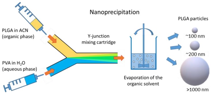
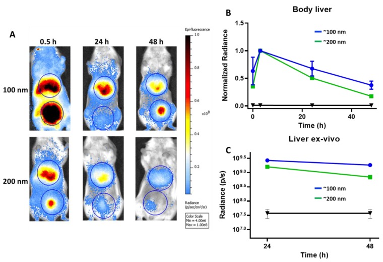
Polymeric particles made up of biodegradable and biocompatible polymers such as poly(lactic-co-glycolic acid) (PLGA) are promising tools for several biomedical applications including drug delivery. Particular emphasis is placed on the size and surface functionality of these systems as they are regarded as the main protagonists in dictating the particle behavior in vitro and in vivo. Current methods of manufacturing polymeric drug carriers offer a wide range of achievable particle sizes, however, they are unlikely to accurately control the size while maintaining the same production method and particle uniformity, as well as final production yield. Microfluidics technology has emerged as an efficient tool to manufacture particles in a highly controllable manner. Here, we report on tuning the size of PLGA particles at diameters ranging from sub-micron to microns using a single microfluidics device, and demonstrate how particle size influences the release characteristics, cellular uptake and in vivo clearance of these particles. Highly controlled production of PLGA particles with ~100 nm, ~200 nm, and >1000 nm diameter is achieved through modification of flow and formulation parameters. Efficiency of particle uptake by dendritic cells and myeloid-derived suppressor cells isolated from mice is strongly correlated with particle size and is most efficient for ~100 nm particles. Particles systemically administered to mice mainly accumulate in liver and ~100 nm particles are cleared slower. Our study shows the direct relation between particle size varied through microfluidics and the pharmacokinetics behavior of particles, which provides a further step towards the establishment of a customizable production process to generate tailor-made nanomedicines.

Keywords: PLGA; drug delivery systems; microfluidics; microparticles; nanoparticles.


Conflict of interest statement

The authors declare no conflict of interest.

Figures

Scheme 1
Scheme 1
Schematic illustration of the particle generation process via nanoprecipitation method using a Y-junction mixing cartridge. PLGA: poly(lactic-co-glycolic acid); ACN: acetonitrile; PVA: polyvinyl alcohol.
Figure 1
Figure 1
(AC) Influence of process and formulation parameters on the particle size. The variation of particle size at different flow rates for 33.3 mg/mL PLGA as organic phase and 1% PVA as aqueous phase. (D) The influence of PLGA concentration on the particle size (organic:aqueous flow rate of 4:4 mL/min, 3% PVA as aqueous phase). Bars: intensity-averaged particle size; green dots: polydispersity index (PDI). QO: flow rate of organic phase, QA: flow rate of aqueous phase. Significant: *: p < 0.05; **: p < 0.005; ***: p < 0.001; ****: p < 0.0001. Non-significant (n.s.): p > 0.05.
Figure 2
Figure 2
Atomic force microscopy height images of (A) >1000 nm, (B), ~200 nm, and (C) ~100 nm PLGA particles. The scale bars display the Z-range and the scan sizes are shown on the x-axis. (D) Size distribution and (E) ζ potential values of PLGA particles measured using dynamic light scattering. (F) Release profile of BODIPY-C12 from >1000 nm, ~200 nm, and ~100 nm particles. Data obtained for >1000 nm, ~200 nm, and ~100 nm particles are represented in orange, blue, and red, respectively.
Figure 3
Figure 3
(A) Intracellular mean fluorescence intensity (MFI) values obtained for >1000 nm (orange), ~200 nm (blue), and ~100 nm (red) PLGA particles incubated with bone marrow-derived dendritic cells (BMDCs) for different time periods, and (B) correlation between the particle size and intracellular MFI after 2 h of incubation with CD103+ dendritic cells, (C) monocytic myeloid-derived suppressor cells (MDSCs), and (D) polymorphonuclear MDSCs isolated from mouse. Data were obtained using flow cytometry.
Figure 4
Figure 4
(A) Fluorescent whole-body images of mice obtained at different time points up to 48 h after intravenous (i.v.) administration of ~200 nm and ~100 nm PLGA particles. The liver and bladder are encircled. (B) Variations of liver fluorescence in whole-body images. The fluorescence intensities are normalized at maximum values observed at 3 h after administration of ~100 nm (blue) and ~200 nm (green) PLGA particles. (C) Variations of liver fluorescence intensities of ~100 nm (blue) and ~200 nm (green) PLGA particles obtained on an isolated liver at 24 h and 48 h after particle administration. Data obtained for untreated mice (negative control) are shown in black.

References

    1. Khalid M., El-Sawy H.S. Polymeric nanoparticles: Promising platform for drug delivery. Int. J. Pharm. 2017;528:675–691. - PubMed
    1. Kumari A., Yadav S.K., Yadav S.C. Biodegradable polymeric nanoparticles based drug delivery systems. Colloids Surf. B Biointerfaces. 2010;75:1–18. doi: 10.1016/j.colsurfb.2009.09.001. - DOI - PubMed
    1. Ud Din F., Aman W., Ullah I., Qureshi O.S., Mustapha O., Shafique S., Zeb A. Effective use of nanocarriers as drug delivery systems for the treatment of selected tumors. Int. J. Nanomed. 2017;12:7291. doi: 10.2147/IJN.S146315. - DOI - PMC - PubMed
    1. Chan J.M., Valencia P.M., Zhang L., Langer R., Farokhzad O.C. Cancer Nanotechnology. Springer; Berlin/Heidelberg, Germany: 2010. Polymeric nanoparticles for drug delivery; pp. 163–175. - PubMed
    1. Hines D.J., Kaplan D.L. Poly (lactic-co-glycolic) acid—controlled-release systems: Experimental and modeling insights. Crit. Rev. Ther. Drug Carr. Syst. 2013;30:257–276. doi: 10.1615/CritRevTherDrugCarrierSyst.2013006475. - DOI - PMC - PubMed

LinkOut - more resources