. 2019 Nov 9;11(11):2716.
doi: 10.3390/nu11112716.

Anti-Osteoporotic Effects of Combined Extract of Lycii Radicis Cortex and Achyranthes japonica in Osteoblast and Osteoclast Cells and Ovariectomized Mice

Affiliations

Anti-Osteoporotic Effects of Combined Extract of Lycii Radicis Cortex and Achyranthes japonica in Osteoblast and Osteoclast Cells and Ovariectomized Mice

Eunkuk Park et al. Nutrients. .

Abstract

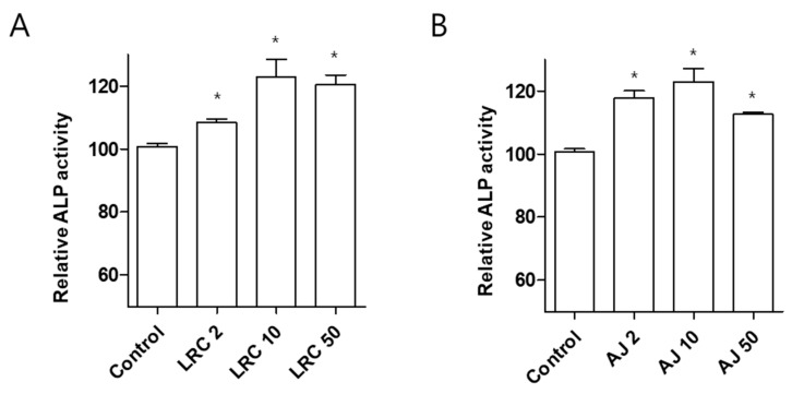
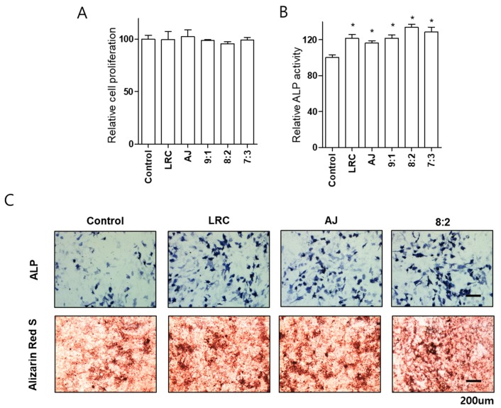
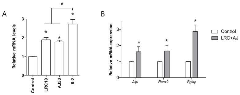
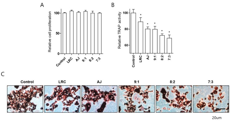
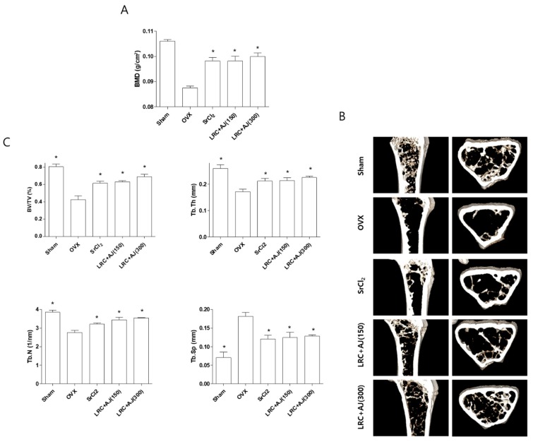
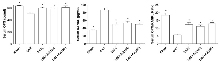
Osteoporosis is characterized by low bone density and quality with high risk of bone fracture. Here, we investigated anti-osteoporotic effects of natural plants (Lycii Radicis Cortex (LRC) and Achyranthes japonica (AJ)) in osteoblast and osteoclast cells in vitro and ovariectomized mice in vivo. Combined LRC and AJ enhanced osteoblast differentiation and mineralized bone-forming osteoblasts by the up-regulation of bone metabolic markers (Alpl, Runx2 and Bglap) in the osteoblastic cell line MC3T3-E1. However, LRC and AJ inhibited osteoclast differentiation of monocytes isolated from mouse bone marrow. In vivo experiments showed that treatment of LRC+AJ extract prevented OVX-induced trabecular bone loss and osteoclastogenesis in an osteoporotic animal model. These results suggest that LRC+AJ extract may be a good therapeutic agent for the treatment and prevention of osteoporotic bone loss.

Keywords: Achyranthes japonica; Lycii Radicis Cortex; osteoblast; osteoclast; osteoporosis; ovariectomized mice.


Conflict of interest statement

The authors declare no conflict of interest.

Figures

Figure 1
Figure 1
Effects of LRC and AJ extracts on cellular differentiation of the osteoblast-lineage cell line MC3T3-E1. Assessment of alkaline phosphatase (ALP) activity is shown for LRC (A) and AJ (B) extracts in MC3T3-E1 osteoblastic cells. After induction of osteoblast differentiation with 50 μg/mL of ascorbic acid and 10 mM of β-glycerophosphate, cells were cultured with three different concentrations (2, 10, and 50 μg/mL) for 3 days, and alkaline phosphatase (ALP) activity was analyzed. *: p < 0.05 vs. Control.
Figure 2
Figure 2
Effects of single or combined LRC and AJ extracts on cellular proliferation, differentiation, and mineralized nodule formation of the osteoblast-lineage cell lines. Assessment is shown for cell proliferation (A) and alkaline phosphatase (ALP) activity (B) of single or combined LRC and AJ (9:1, 8:2 or 7:3 ratio) in MC3T3-E1 cells. Osteoblast differentiation was induced by adding 50 μg/mL of ascorbic acid and 10 mM of β-glycerophosphate, MC3T3-E1 cells were incubated with 10 μg/mL of single or combined LRC and AJ extracts (9:1, 8:2 or 7:3) for 3 days, and then cellular proliferation and ALP activity were assessed. (C) ALP and Alizarin Red S staining was assessed in single or combined LRC and AJ-treated cells. Cells were treated with 10 μg/mL of single or combined LRC and AJ extracts (8:2) for 3 days (for ALP staining) or 21 days (for Alizarin Red S staining), and osteoclast differentiation and mineralized nodule formation were stained. Control: non-treated cells. *: p < 0.05 vs. Control.
Figure 3
Figure 3
Effects of LRC and AJ extract on mRNA expression of bone formation markers (Alpl, Runx2 and Bglap) in MC3T3-E1 cells. (A) Cells were treated with single extracts of LRC and AJ and combined LRC and AJ, with an 8:2 ratio at 10 μg/mL. Total RNA of the cells was extracted and then the mRNA expression level of Bglap was compared by quantitative reverse-transcription polymerase chain reaction (qRT-PCR). (B) The mRNA expression of Alpl, Runx2 and Bglap was confirmed in the combined LRC and AJ extracts. The mRNA levels of bone formation markers were normalized by mRNA expression of Gapdh (glyceraldehyde 3-phosphate dehydrogenase). Control: non-treated cells. *: p < 0.05 vs. Control and #: p < 0.05 vs. 8:2.
Figure 4
Figure 4
Effects of single or combined LRC and AJ extracts on cellular proliferation and differentiation of primary-cultured monocytes. Assessment were made of cell proliferation (A) and tartrate-resistant acid phosphatase (TRAP) activity in monocytes from mouse bone marrow (B). Osteoclast differentiation was induced by adding 30 ng/mL of M-CSF and 50 ng/mL of RANKL, and cells were treated with single or combined LRC and AJ (9:1, 8:2 or 7:3 ratio). The differentiated osteoclast cells were analyzed by TRAP activity (B) and TRAP staining (C). Control: Non-treated monocyte cells cultured with M-CSF and RANKL. *: p < 0.05 vs. Control.
Figure 5
Figure 5
Anti-osteoporotic effects of LRC+AJ extract on the improvement of bone mineral density (BMD) and bone structural properties in OVX mice. The OVX mice were administered with either SrCl2 (10 mg/kg/day) or LRC+AJ extract (150 or 300 mg/kg/day) for 12 weeks. (A) BMD of the right femur was measured using a PIXI-mus bone densitometer. (B) Transverse micro-CT images were scanned using microcomputed tomography (CT). (C) Trabecular bone structural properties of bone volume (BV/TV), trabecular thickness (Tb.Th), number (Tb.N) and spacing (Tb.Sp) were analyzed at the end of the experiment. Sham: sham operated, OVX: non-Scopolin-administered mice. *: p < 0.05 vs. OVX group.
Figure 6
Figure 6
Effects of LRC+AJ extract on serum levels of bone metabolic makers (osteoprotegrin (OPG) and Receptor Activator of Nuclear Factor kappa-B ligand (RANKL)). After 12 weeks of administration, the blood samples were collected and serum levels of OPG and RANKL were measured using ELISA. *: p < 0.05 vs. OVX control.

References

    1. Feng X., McDonald J.M. Disorders of bone remodeling. Annu. Rev. Pathol. 2011;6:121–145. doi: 10.1146/annurev-pathol-011110-130203. - DOI - PMC - PubMed
    1. Raggatt L.J., Partridge N.C. Cellular and molecular mechanisms of bone remodeling. J. Biol. Chem. 2010;285:25103–25108. doi: 10.1074/jbc.R109.041087. - DOI - PMC - PubMed
    1. Zheng M., Ge Y., Li H., Yan M., Zhou J., Zhang Y. Bergapten prevents lipopolysaccharide mediated osteoclast formation, bone resorption and osteoclast survival. Int. Orthop. 2014;38:627–634. doi: 10.1007/s00264-013-2184-y. - DOI - PMC - PubMed
    1. Moreira L.D., Oliveira M.L., Lirani-Galvao A.P., Marin-Mio R.V., Santos R.N., Lazaretti-Castro M. Physical exercise and osteoporosis: Effects of different types of exercises on bone and physical function of postmenopausal women. Arq. Bras. Endocrinol. Metabol. 2014;58:514–522. doi: 10.1590/0004-2730000003374. - DOI - PubMed
    1. Bernhardt A., Schamel M., Gbureck U., Gelinsky M. Osteoclastic differentiation and resorption is modulated by bioactive metal ions Co2+, Cu2+ and Cr3+ incorporated into calcium phosphate bone cements. PLoS ONE. 2017;12:e0182109. doi: 10.1371/journal.pone.0182109. - DOI - PMC - PubMed

LinkOut - more resources