. 2019 Nov 9;11(11):1766.
doi: 10.3390/cancers11111766.

Collagen Induces a More Proliferative, Migratory and Chemoresistant Phenotype in Head and Neck Cancer via DDR1

Affiliations

Collagen Induces a More Proliferative, Migratory and Chemoresistant Phenotype in Head and Neck Cancer via DDR1

Sook Ling Lai et al. Cancers (Basel). .

Abstract

Head and neck squamous cell carcinoma (HNSCC) is the sixth most common cancer worldwide and includes squamous cell carcinomas of the oropharynx and oral cavity. Patient prognosis has remained poor for decades and molecular targeted therapies are not in routine use. Here we showed that the overall expression of collagen subunit genes was higher in cancer-associated fibroblasts (CAFs) than normal fibroblasts. Focusing on collagen8A1 and collagen11A1, we showed that collagen is produced by both CAFs and tumour cells, indicating that HNSCCs are collagen-rich environments. We then focused on discoidin domain receptor 1 (DDR1), a collagen-activated receptor tyrosine kinase, and showed that it is over-expressed in HNSCC tissues. Further, we demonstrated that collagen promoted the proliferation and migration of HNSCC cells and attenuated the apoptotic response to cisplatin. Knockdown of DDR1 in HNSCC cells demonstrated that these tumour-promoting effects of collagen are mediated by DDR1. Our data suggest that specific inhibitors of DDR1 might provide novel therapeutic opportunities to treat HNSCC.

Keywords: DDR1; collagen; head and neck cancer.


Conflict of interest statement

The authors declare no conflict of interest. The funders had no role in the design of the study; in the collection, analyses, or interpretation of data; in the writing of the manuscript; or in the decision to publish the results.

Figures

Figure 1
Figure 1
Collagen expression in cancer-associated fibroblasts (CAFs) and head and neck squamous cell carcinoma (HNSCC) cells. (A) Extracellular structure organization was the most significantly enriched gene ontology (GO) term identified by gene set enrichment analysis (GSEA) of genes differentially expressed between normal human oral fibroblasts (NHOFs) and CAFs, as determined by RNAseq. (B) Heatmap highlighting the elevated expression of collagen subtypes in CAFs. (C) Total collagen expression (average expression of all collagen subtypes from RNAseq) was higher in the majority of CAF strains compared to normal fibroblasts. (D) Expression of COL8A1 and COL11A1 in NHOFs and CAFs, as determined by RNAseq. (E) RT-qPCR was used to confirm the expression of COL8A1 and COL11A1 in fibroblast strains. (F) RT-qPCR analysis showed COL8A1 and COL11A1 were also expressed in some HNSCC cell lines. Data for the RT-qPCR experiments are shown as mean +/− SD values of triplicates.
Figure 2
Figure 2
Expression of COL8A1 and COL11A1 in oropharyngeal squamous cell carcinoma (OPSCC). Photomicrographs show expression of COL8A1 (top panel) and COL11A1 (lower panel) in OPSCC tissues. Tissues were multiplex-stained with COL8A1 or COL11A1 (fluorescein, green), together with α-smooth muscle actin (α-SMA) (Cy3, red) antibodies and 4′,6-diamidino-2-phenylindole (DAPI) (blue). The expression of COL8A1and COL11A1 was detected in OPSCCs and cancer-associated fibroblasts. Representative images are shown and were captured with a confocal laser microscope (Zeiss, Carl Zeiss AG, Oberkochen, Germany; magnification × 63). Examples of COL8A1and COL11A1 expression in oral squamous cell carcinoma tissues are shown in Supplementary Figure S2.
Figure 3
Figure 3
Discoidin domain receptor 1 (DDR1) was over-expressed in head and neck squamous cell carcinoma (HNSCC). (A) DDR1 is readily detectable in HNSCC cell lines by RT-qPCR and western blotting. (B) Analysis of The Cancer Genome Atlas (TCGA) expression data revealed that DDR1 is significantly over-expressed in tumours relative to normal samples. There was no statistically significant difference in DDR1 expression between human papillomavirus (HPV)-negative and HPV-positive tumours. (C) Immunohistochemical analysis of DDR1 protein revealed that normal epithelium showed weak cytoplasmic staining (i and ii), whilst the majority of squamous cell carcinomas (8 of 12) showed increased DDR1 expression in comparison to normal epithelium (iii and iv). (Original magnification × 100).
Figure 4
Figure 4
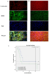
Expression of discoidin domain receptor 1 (DDR1) in oropharyngeal squamous cell carcinoma (OPSCC). Tissues were multiplex-stained with pan-cytokeratin cocktail AE1/AE3 (Cy3, red) and DDR1 (fluorescein, green) antibodies, plus 4′,6-diamidino-2-phenylindole (DAPI) (blue) nuclear counterstain. DDR1 expression in OPSCCs was (A) cytoplasmic and membraneous or (B) membraneous. Representative images are shown and were captured using Metamorph Pathology Imaging System (Nikon, Tokyo, Japan; magnification ×60). Examples of DDR1 expression in oral squamous cell carcinoma tissues are shown in Supplementary Figure S5. (C) High DDR1 expression in OPSCC patients was correlated with worse survival. Patients with high DDR1 expression have a lower 5-year survival rate (33%) than that of patients with low DDR1 expression (78%), log-rank (Mantel–Cox) (p = 0.022).
Figure 5
Figure 5
Collagen promoted the proliferation and migration of head and neck squamous cell carcinoma (HNSCC) cells and suppressed the response to cisplatin. (A) Collagen significantly increased the growth of the HNSCC cell lines, SCC040, SCC154, VU040T and VU147T. (B) Pre-treatment of cells with collagen significantly enhanced the migration of cells through fibronectin-coated membranes. (C) Cells pre-treated with collagen were significantly less sensitive to cisplatin, as determined by Annexin V-FITC/Propidium Iodide staining following treatment with 50 μM cisplatin for 72 hours. Results shown are mean +/− standard deviation values of triplicates. *, **, ***, **** denote p < 0.05, p < 0.01, p < 0.001 and p < 0.0001 respectively.
Figure 6
Figure 6
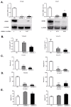
The effects of collagen are mediated by discoidin domain receptor 1 (DDR1). (A) Knockdown of DDR1 following stable transduction of VU040 and VU147T cells with two independent short hairpin RNAs. RT-qPCR showed a marked reduction in DDR1 mRNA (top panel) and total DDR1 protein levels (bottom panel). The expression of DDR1 mRNA in NS (control) cells was normalized to 1. (B) Following DDR1 knockdown, VU040 and VU147T cells grew slower compared to NS controls in the presence of collagen. (C) Knockdown of DDR1 inhibited the migration of VU040T and VU147T cells pre-treated with collagen in Transwell assays. (D) Knockdown of DDR1 inhibited the invasion of VU040T and VU147T cells through matrigel-coated filters in Transwell assays. (E) The protective effects of collagen on cisplatin-induced apoptosis were reversed following DDR1 knockdown. Results shown are mean +/- standard deviation values of triplicates. *, **, *** denotes p < 0.05, p < 0.01 and p < 0.001 and p < 0.0001 respectively.

References

    1. Warnakulasuriya S. Global epidemiology of oral and oropharyngeal cancer. Oral Oncol. 2009;45:309–316. doi: 10.1016/j.oraloncology.2008.06.002. - DOI - PubMed
    1. Speight P., Farthing P. The pathology of oral cancer. Br. Dent. J. 2018;225:841. doi: 10.1038/sj.bdj.2018.926. - DOI - PubMed
    1. Mehanna H., Beech T., Nicholson T., El-Hariry I., McConkey C., Paleri V., Roberts S. Prevalence of human papillomavirus in oropharyngeal and nonoropharyngeal head and neck cancer—Systematic review and meta-analysis of trends by time and region. Head Neck. 2013;35:747–755. doi: 10.1002/hed.22015. - DOI - PubMed
    1. Leemans C.R., Snijders P.J., Brakenhoff R.H. The molecular landscape of head and neck cancer. Nat. Rev. Cancer. 2018;18:269–282. doi: 10.1038/nrc.2018.11. - DOI - PubMed
    1. Prime S., Cirillo N., Hassona Y., Lambert D., Paterson I., Mellone M., Thomas G., James E., Parkinson E. Fibroblast activation and senescence in oral cancer. J. Oral Pathol. Med. 2017;46:82–88. doi: 10.1111/jop.12456. - DOI - PubMed