. 2019 Oct 25;8(11):1322.
doi: 10.3390/cells8111322.

Perturbation of Methionine/S-adenosylmethionine Metabolism as a Novel Vulnerability in MLL Rearranged Leukemia

Affiliations

Perturbation of Methionine/S-adenosylmethionine Metabolism as a Novel Vulnerability in MLL Rearranged Leukemia

Aditya Barve et al. Cells. .

Abstract

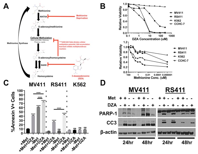
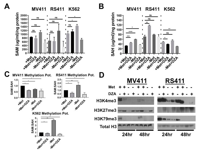
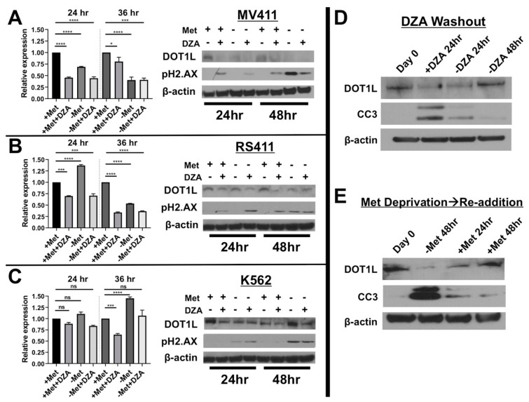
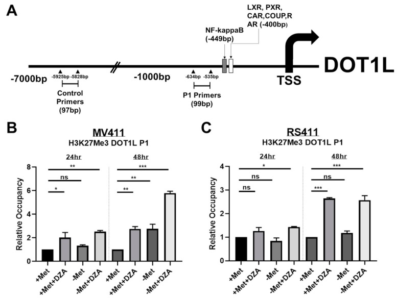
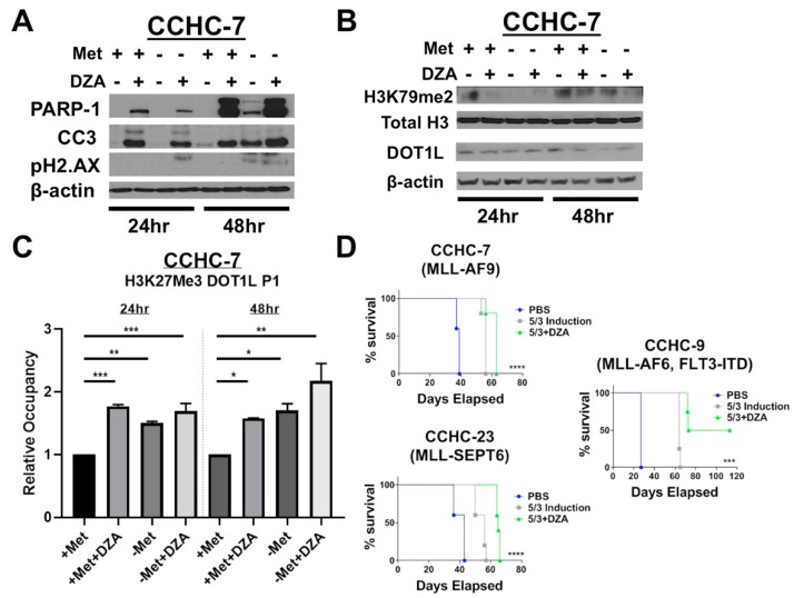
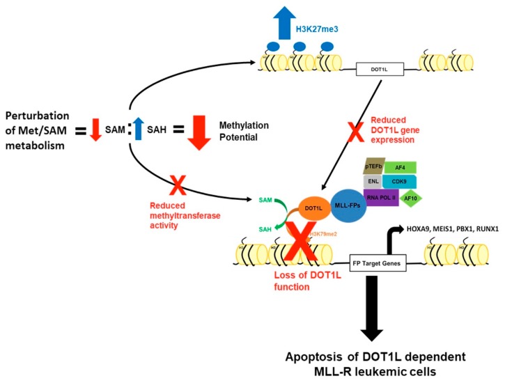
Leukemias bearing mixed lineage leukemia (MLL) rearrangement (MLL-R) resulting in expression of oncogenic MLL fusion proteins (MLL-FPs) represent an especially aggressive disease subtype with the worst overall prognoses and chemotherapeutic response. MLL-R leukemias are uniquely dependent on the epigenetic function of the H3K79 methyltransferase DOT1L, which is misdirected by MLL-FPs activating gene expression, driving transformation and leukemogenesis. Given the functional necessity of these leukemias to maintain adequate methylation potential allowing aberrant activating histone methylation to proceed, driving leukemic gene expression, we investigated perturbation of methionine (Met)/S-adenosylmethionine (SAM) metabolism as a novel therapeutic paradigm for MLL-R leukemia. Disruption of Met/SAM metabolism, by either methionine deprivation or pharmacologic inhibition of downstream metabolism, reduced overall cellular methylation potential, reduced relative cell numbers, and induced apoptosis selectively in established MLL-AF4 cell lines or MLL-AF6-expressing patient blasts but not in BCR-ABL-driven K562 cells. Global histone methylation dynamics were altered, with a profound loss of requisite H3K79 methylation, indicating inhibition of DOT1L function. Relative occupancy of the repressive H3K27me3 modification was increased at the DOT1L promoter in MLL-R cells, and DOT1L mRNA and protein expression was reduced. Finally, pharmacologic inhibition of Met/SAM metabolism significantly prolonged survival in an advanced, clinically relevant patient-derived MLL-R leukemia xenograft model, in combination with cytotoxic induction chemotherapy. Our findings provide support for further investigation into the development of highly specific allosteric inhibitors of enzymatic mediators of Met/SAM metabolism or dietary manipulation of methionine levels. Such inhibitors may lead to enhanced treatment outcomes for MLL-R leukemia, along with cytotoxic chemotherapy or DOT1L inhibitors.

Keywords: AML; MLL; Methionine; SAM; mouse model.


Conflict of interest statement

The authors declare no conflict of interest.

Figures

Figure 1
Figure 1
Perturbation of methionine (Met)/S-adenosylmethionine (SAM) metabolism potently induces apoptosis and reduces cell viability in established mixed lineage leukemia rearrangement (MLL-R) leukemia cell lines. (A) Simplified schematic of Met/SAM metabolism showing important enzymatic mediators, substrates and products. Deregulation of Met/SAM metabolism and subsequently methylation potential was achieved by targeting two distinct nodes, either by reducing synthesis of the methyl donor moiety SAM by methionine deprivation or pharmacological inhibition of downstream SAH metabolism and methionine recycling. (B) Dose and time dependent reduction in relative survival of MLL-AF4 expressing MV411 (AML) and RS411 (ALL) cells treated with increasing concentrations 3-deazaadenosine (DZA) as quantified by Alamar Blue vitality assay 48 h post treatment. K562 cells lacking MLL-fusion expression show no reduction in relative survival upon DZA treatment (left). MLL-R cells also show a greater reduction in cell numbers upon methionine withdrawal as compared to K562 cells (right). (C) From left to right; Methionine deprivation or exposure to 15µM DZA potently induced apoptosis at 48 h in MV411 cells singly, and the combination showed an additive increase in apoptosis as determined by Annexin V+ staining. RS411 cells only undergo apoptosis upon exposure to 15µM DZA, and this effect is amplified by methionine deprivation. K562 cells lacking MLL-R show no induction of apoptosis 48 h post DZA exposure or methionine deprivation (Tukey HSD, p < 0.0001). (D) Changes in protein expression corresponding with apoptosis induction (PARP-1 and Caspase-3 cleavage) were observed under all experimental conditions in MV411 cells (left), while RS411 cells only undergo apoptosis upon DZA-mediated SAHH inhibition (right).
Figure 2
Figure 2
Disruption of Met/SAM metabolism increases intracellular SAH concentration and decreases overall methylation potential. (A) From left to right; Intracellular SAM concentration was not significantly changed in either MV411 or RS411 cells but was significantly elevated in K562 cells upon methionine starvation and significantly reduced with DZA treatment and methionine deprivation (Tukey HSD, * = p < 0.05). (B) From left to right; Intracellular SAH concentration was increased in MV411 cells under all experimental conditions and timepoints, while only DZA-mediated inhibition of SAH metabolism sufficiently increased intracellular SAH concentration in RS411 cells (Tukey HSD, *= p < 0.05 ** = p < 0.01, *** = p < 0.001, **** = p < 0.0001). (C) Overall cellular methylation potential (SAM:SAH) was significantly reduced in MV411 cells 24 h post exposure to experimental conditions. RS411 cells only show significant reduction in methylation potential following 24 h exposure to DZA, and conversely, a significant increase in methylation potential was observed following 24 h of methionine deprivation (Tukey HSD, * = p <0.05 ** = p <0.01, *** = p <0.001, **** = p <0.0001). (D) Global histone methylation dynamics are altered in both MLL-AF4 expressing cell lines upon disruption of Met/SAM metabolism resulting in a reduction in the activating H3K4me3 modification and an increase in the repressive H3K27me3 following DZA-mediated SAHH inhibition. Importantly, perturbation of Met/SAM metabolism potently reduced global levels of DOT1L-dependent H3K79me2, an activating modification absolutely required by MLL-R leukemic cells.
Figure 3
Figure 3
Alteration of Met/SAM metabolism reduces mRNA and protein expression of the required H3K79 methyltransferase DOT1L selectively in MLL-R leukemia cells. (A) DZA treatment or methionine deprivation of MV411 cells significantly reduced DOT1L mRNA (Tukey HSD, * = p < 0.05 *** = p <0.001 **** = p <0.0001) following 24 or 36 h exposure to experimental conditions (left) and these changes are reflected in DOT1L protein levels (right). Reduction in DOT1L expression also correlated with the induction of DNA damage as determined by pH2.AX formation, likely due to the critical role DOT1L plays in regulation of the DNA damage repair response. (B) RS411 cells display a significant reduction in DOT1L mRNA (Tukey HSD, *** = p <0.001 **** = p< 0.0001) and protein levels (right) following DZA mediated SAHH inhibition, but not with methionine deprivation alone at 24 h. By 36 h methionine deprivation alone significantly reduced DOT1L mRNA (Tukey HSD, **** = p< 0.0001) and protein expression in RS411 cells, and again DNA damage induction was detected only under conditions of DOT1L reduction. (C) Non-MLL-R K562 cells lacking functional dependence on DOT1L show no significant changes in DOT1L mRNA expression 24 h post study initiation, however DOT1L mRNA was significantly reduced 36 h after DZA exposure, but was significantly increased following 36 h of methionine deprivation (Tukey HSD, *** = p < 0.001 **** = p < 0.0001). Interestingly, modulation of DOT1L protein levels was observed even in K562 cells (right), and pH2.AX foci formation was also detected only under conditions of methionine deprivation in K562 cells. These results may suggest that appropriate regulation of Met/SAM metabolism and one-carbon sensing may directly play a more universal role in regulation of DOT1L expression and function in multiple cell types, regardless of MLL-R mediated functional dependence on DOT1L and H3K79 methylation. (D) To further correlate SAM/Met metabolism with DOT1L expression RS411 cells were cultured for 24 h in the presence of DZA after which cells were washed and replated in DZA free, methionine containing media. Total protein lysates were collected at the indicated timepoints, and immunoblotting revealed that DOT1L expression was decreased following DZA treatment with an increase in caspase-3 cleavage. Washout of DZA increased DOT1L expression back to initial levels, with a corresponding reduction in cleaved caspase-3. (E) MV411 cells deprived of methionine for 48 h had reduced DOT1L protein expression and increased caspase-3 cleavage. Reintroduction of methionine in the media rescued DOT1L expression to initial levels, with a corresponding reduction in caspase-3 cleavage.
Figure 4
Figure 4
Decreased expression of DOT1L is correlated to changes in histone methylation dynamics specifically at the DOT1L promoter. (A) DOT1L promoter map illustrating putative transcription factor binding sites as determined by MotifMap software, as well as the specific binding sites of the DOT1L promoter specific primers (P1) and the distal control primers. (B) MV411 cells show significantly elevated levels of H3K27me3 occupancy at the DOT1L promoter following DZA treatment or methionine deprivation and by 48 h an additive increase was observed under simultaneous methionine deprivation and DZA-mediated SAHH inhibition (Tukey HSD, *= p <0.05 **= p <0.01 ***= p <0.001). (C) RS411 cells exhibit significantly increased DOT1L promoter H3K27me3 occupancy following 24 h of simultaneous methionine deprivation and DZA exposure, and by 48 h, DZA treatment alone but not methionine deprivation alone is sufficient to significantly elevate DOT1L-promoter H3K27me3 occupancy (Tukey HSD, *= p <0.05 ***= p <0.001).
Figure 5
Figure 5
Patient-derived MLL-R leukemic blasts are sensitive to perturbation of Met/SAM metabolism and show changes in global and DOT1L promoter–specific histone methylation dynamics consistent with tested MLL-R cell lines. Combining DZA with 5 + 3 induction therapy significantly prolonged the life of mice harboring several different MLL-R patient derived xenografts. (A) Patient-derived CCHC-7 cells behave similarly to the RS411 cell line and only undergo apoptosis following DZA-mediated SAHH inhibition with or without methionine deprivation, as determined by PARP-1 and caspase-3 cleavage. Methionine deprivation for 48 h was also sufficient to induce apoptosis and PARP-1 and caspase-3 cleavage. (B) CCHC-7 cells show a profound early loss in global H3K79me2 levels following 8 h of exposure to experimental conditions, and this reduction was more modest 24 h post exposure to either DZA or methionine deprivation alone. However, the combination of simultaneous DZA mediated SAHH inhibition and methionine deprivation was still sufficient to decrease global H3K79me2 even after 24 h (top). DOT1L protein expression was also reduced in CCHC-7 cells by methionine deprivation and/or DZA treatment (bottom). (C) CCHC-7 cells display significantly elevated levels of DOT1L promoter H3K27me3 occupancy 24 or 48 h after exposure to all experimental conditions (Tukey HSD, *= p <0.05 **= p <0.01 ***= p <0.001). (D) NRGS mice (n = 5 for all cohorts in all studies) were xenografted with 1.25 × 10⁵ CCHC-7, CCHC-23, or CCHC-9 patient leukemic cells each bearing a unique MLL-R, and seven days post xenotransplant, mice began bolus intravenous infusion of isovolumetric amounts of 5 + 3 induction therapy (3 mg/kg doxorubicin, days 1–3 and 75 mg/kg cytarabine, Days 1–5), 5 + 3 induction plus 25 mg/kg DZA (days 1–5), or the vehicle PBS alone. DZA in combination with 5 + 3 induction significantly prolonged the life of these mice as compared to either 5 + 3 induction therapy or vehicle treatment alone in all xenograft studies regardless of particular patient MLL-R (Mantel-Cox, *** = p <0.001 *** *= p <0.0001), and promisingly, two of the five mice bearing CCHC-9 xenografts completely failed to develop leukemic disease and survived till the endpoint of our study (far right).
Figure 6
Figure 6
A mechanism by which perturbation of Met/SAM metabolism induces cytotoxicity and apoptosis in MLL-R-bearing cells. Perturbation of Met/SAM metabolism, either by deprivation of exogenous methionine or by pharmacologic inhibition of downstream SAH metabolism, results in a significant increase in the levels of intracellular SAH leading to an overall decrease in global cellular methylation potential. Under conditions of decreased methylation potential, there is a global reduction in methyltransferase activity and a DOT1L promoter–specific increase in the repressive H3K27me3 modification, resulting in decreased DOT1L expression and function. MLL-R leukemic cells are uniquely dependent on DOT1L expression and function to maintain aberrant leukemogenic gene expression and survival, and loss of DOT1L function and expression may partially explain their increased sensitivity to alterations of Met/SAM metabolism.

References

    1. Winters A.C., Bernt K.M. MLL-Rearranged Leukemias-An Update on Science and Clinical Approaches. Front. Pediatr. 2017;5:4. doi: 10.3389/fped.2017.00004. - DOI - PMC - PubMed
    1. Biondi A., Cimino G., Pieters R., Pui C.H. Biological and therapeutic aspects of infant leukemia. Blood. 2000;96:24–33. doi: 10.1182/blood.V96.1.24. - DOI - PubMed
    1. Bernt K.M., Armstrong S.A. Targeting epigenetic programs in MLL-rearranged leukemias. Hematol. Am. Soc. Hematol. Educ. Program. 2011;2011:354–360. doi: 10.1182/asheducation-2011.1.354. - DOI - PubMed
    1. Krivtsov A.V., Armstrong S.A. MLL translocations, histone modifications and leukaemia stem-cell development. Nat. Rev. Cancer. 2007;7:823–833. doi: 10.1038/nrc2253. - DOI - PubMed
    1. Okada Y., Feng Q., Lin Y., Jiang Q., Li Y., Coffield V.M., Su L., Xu G., Zhang Y. hDOT1L links histone methylation to leukemogenesis. Cell. 2005;121:167–178. doi: 10.1016/j.cell.2005.02.020. - DOI - PubMed

Publication types

MeSH terms

LinkOut - more resources