. 2019 Oct 26;11(11):1656.
doi: 10.3390/cancers11111656.

High Clinical Value of Liquid Biopsy to Detect Circulating Tumor Cells and Tumor Exosomes in Pancreatic Ductal Adenocarcinoma Patients Eligible for Up-Front Surgery

Affiliations

High Clinical Value of Liquid Biopsy to Detect Circulating Tumor Cells and Tumor Exosomes in Pancreatic Ductal Adenocarcinoma Patients Eligible for Up-Front Surgery

Etienne Buscail et al. Cancers (Basel). .

Abstract

Purpose: Expediting the diagnosis of pancreatic ductal adenocarcinoma (PDAC) would benefit care management, especially for the start of treatments requiring histological evidence. This study evaluated the combined diagnostic performance of circulating biomarkers obtained by peripheral and portal blood liquid biopsy in patients with resectable PDAC.

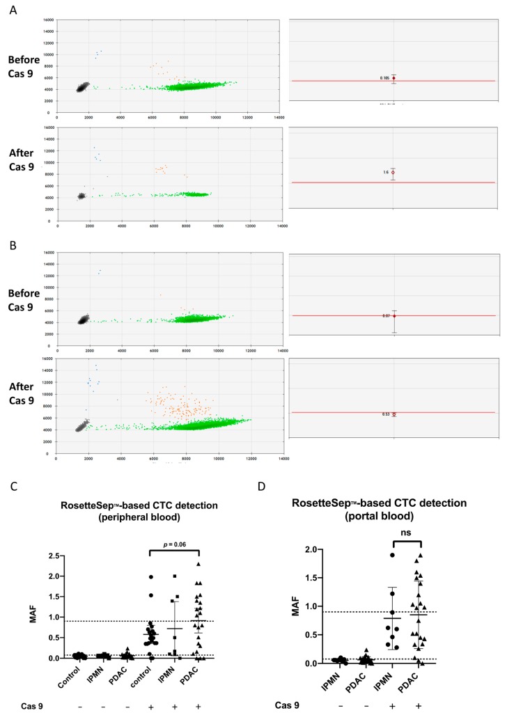
Experimental design: Liquid biopsies were performed in a prospective translational clinical trial (PANC-CTC #NCT03032913) including 22 patients with resectable PDAC and 28 noncancer controls from February to November 2017. Circulating tumor cells (CTCs) were detected using the CellSearch® method or after RosetteSep® enrichment combined with CRISPR/Cas9-improved KRAS mutant alleles quantification by droplet digital PCR. CD63 bead-coupled Glypican-1 (GPC1)-positive exosomes were quantified by flow cytometry.

Results: Liquid biopsies were positive in 7/22 (32%), 13/22 (59%), and 14/22 (64%) patients with CellSearch® or RosetteSep®-based CTC detection or GPC1-positive exosomes, respectively, in peripheral and/or portal blood. Liquid biopsy performance was improved in portal blood only with CellSearch®, reaching 45% of PDAC identification (5/11) versus 10% (2/22) in peripheral blood. Importantly, combining CTC and GPC1-positive-exosome detection displayed 100% of sensitivity and 80% of specificity, with a negative predictive value of 100%. High levels of GPC1+-exosomes and/or CTC presence were significantly correlated with progression-free survival and with overall survival when CTC clusters were found.

Conclusion: This study is the first to evaluate combined CTC and exosome detection to diagnose resectable pancreatic cancers. Liquid biopsy combining several biomarkers could provide a rapid, reliable, noninvasive decision-making tool in early, potentially curable pancreatic cancer. Moreover, the prognostic value could select patients eligible for neoadjuvant treatment before surgery. This exploratory study deserves further validation.

Keywords: circulating tumor cells; exosomes; liquid biopsy; pancreatic cancer.


Conflict of interest statement

The authors declare no conflict of interest.

Figures

Figure 1
Figure 1
Study design, blood samples, and liquid biopsy methods. (A) Pancreatic ductal adenocarcinoma (PDAC) patients and patients with IPMN had both peripheral and portal samples for CTC-enrichment detection/count and quantification of GPC1-positive exosomes (blue rectangle and arrows). (B) Control group had peripheral samples for CTC-enrichment detection (RosetteSepTM) and quantification of GPC1-positive exosomes (green rectangle and arrows). Abbreviations: EVs: extracellular vesicles; CTC: circulating tumor cell; IPMN: intraductal papillary and mucinous neoplasm; GPC1: Glypican 1.
Figure 2
Figure 2
Heat maps of liquid biopsy results. (A) PDAC patients, (B) IPMN patients, and (C) noncancer control individuals. White rectangle: negative result, blue rectangle: positive result, crossed rectangle: not done. In the PDAC heat map, the bottom ladder indicates adenocarcinoma stage rankings from 1 to 3 according the stage of the disease (i.e., stage 1 light blue, stage 2 blue, stage 3 dark blue). In the IPMN heat map, the bottom ladder indicates dysplasia ranking from 0 (white box) for low grade dysplasia to 1 for high grade dysplasia (blue box). PDAC, pancreatic ductal adenocarcinoma; IPMN, intraductal papillary and mucinous neoplasm. (DF) Venn diagrams recapitulating rates of CTC detection by CellSearch® or RosetteSepTM-based enrichment and GPC1-positive-exosome quantification of (D) peripheral blood samples, (E) portal blood samples, (F) combined peripheral and portal blood samples.
Figure 3
Figure 3
Analysis of GPC1-positive-exosome quantification and CellSearch® positive CTC count and clusters according to clinical criteria. Kaplan–Meier curves, with p values (log Rank) for comparison between (A) overall survival (OS) for patients with >20% GPC1-positive exosomes (4 times the median value) and/or with CTC clusters and patient with <20% GPC1-positive exosomes and/or CellSearch® without CTC clusters. (B) Progression-free survival (PFS) for patients with GPC1-positive exosomes and/or CellSearch® positive and GPC1-negative exosomes and/or CellSearch® negative in peripheral blood. (C) Immunofluorescent staining image of captured CTC clusters. Circulating tumor cell clusters captured from a portal vein sample using the CellSearch system. (CK, cytokeratin; PE, phycoerythrin; DAPI, 4′,6-diamidino-2-phenylindole; DAPI stain is purple and CK stain is green, (original magnification ×400)).
Figure 4
Figure 4
The ddPCR results for KRAS detection after CTC enrichment. (A,B) Individual droplet PCR fluorescence results are plotted as two-dimensional dot plots (left). Grey dots correspond to empty droplets. Green dots correspond to droplets containing wild-type (WT) copies of KRAS. Blue dots correspond to droplets containing one mutant KRAS allele. Orange dots correspond to droplets containing WT (X-axis of the left panels corresponding to the HEX, hexachlorofluorescein succinimidyl ester fluorophore) and mutant alleles (Y-axis of the left panels corresponding to the FAM, 6-carboxyfluoresceine fluorophore). On the right panels, MAFs are shown for individual results, with the maximum and the minimum values of triplicates; the red lines indicate the positivity threshold. Patient #36 (A) became positive and patient #39 (B) was negative for KRAS mutation before and after Cas9. (C,D) MAF of KRAS mutation by ddPCR after RosetteSepTM CTC enrichment. Greater median MAF in CTC-enriched samples after CRISPR/Cas9 cut of the wild-type KRAS allele as compared to uncut DNA in (C) peripheral and (D) portal blood. Higher median MAFs in patients compared with the control group tended toward significance (p = 0.06 by Mann–Whitney test). MAF: mutant allele frequency.

References

    1. Buscail L. Commentary: Pancreatic cancer: Is the worst to come? Int. J. Epidemiol. 2017;46:1774–1775. doi: 10.1093/ije/dyx143. - DOI - PubMed
    1. Tempero M.A., Malafa M.P., Al-Hawary M., Asbun H., Bain A., Behrman S.W., Benson A.B., Binder E., Cardin D.B., Cha C., et al. Pancreatic Adenocarcinoma, Version 2.2017, NCCN Clinical Practice Guidelines in Oncology. J. Natl. Compr. Cancer Netw. JNCCN. 2017;15:1028–1061. doi: 10.6004/jnccn.2017.0131. - DOI - PubMed
    1. Zhou B., Xu J.-W., Cheng Y.-G., Gao J.-Y., Hu S.-Y., Wang L., Zhan H.-X. Early detection of pancreatic cancer: Where are we now and where are we going? Int. J. Cancer. 2017;141:231–241. doi: 10.1002/ijc.30670. - DOI - PubMed
    1. Storm A.C., Lee L.S. Endoscopic ultrasound-guided techniques for diagnosing pancreatic mass lesions: Can we do better? World J. Gastroenterol. 2016;22:8658–8669. doi: 10.3748/wjg.v22.i39.8658. - DOI - PMC - PubMed
    1. Bhat K., Wang F., Ma Q., Li Q., Mallik S., Hsieh T.-C., Wu E. Advances in biomarker research for pancreatic cancer. Curr. Pharm. Des. 2012;18:2439–2451. doi: 10.2174/13816128112092439. - DOI - PMC - PubMed

LinkOut - more resources