Review
. 2019 Oct 26;20(21):5337.
doi: 10.3390/ijms20215337.

Insight into Mechanobiology: How Stem Cells Feel Mechanical Forces and Orchestrate Biological Functions

Affiliations
Review

Insight into Mechanobiology: How Stem Cells Feel Mechanical Forces and Orchestrate Biological Functions

Chiara Argentati et al. Int J Mol Sci. .

Abstract

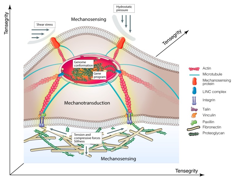
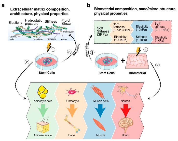
The cross-talk between stem cells and their microenvironment has been shown to have a direct impact on stem cells' decisions about proliferation, growth, migration, and differentiation. It is well known that stem cells, tissues, organs, and whole organisms change their internal architecture and composition in response to external physical stimuli, thanks to cells' ability to sense mechanical signals and elicit selected biological functions. Likewise, stem cells play an active role in governing the composition and the architecture of their microenvironment. Is now being documented that, thanks to this dynamic relationship, stemness identity and stem cell functions are maintained. In this work, we review the current knowledge in mechanobiology on stem cells. We start with the description of theoretical basis of mechanobiology, continue with the effects of mechanical cues on stem cells, development, pathology, and regenerative medicine, and emphasize the contribution in the field of the development of ex-vivo mechanobiology modelling and computational tools, which allow for evaluating the role of forces on stem cell biology.

Keywords: computational tools; ex-vivo stem cell models; mechanosensing; mechanotransduction; regenerative medicine; stem cell-biomaterial interaction; stem cells.


Conflict of interest statement

The authors declare no conflict of interest.

Figures

Figure 1
Figure 1
Schematic illustration of molecular basis of mechanobiology. Cartoon shows how mechanical cues are transmitted to the nucleus via integrins > focal adhesion complex > cytoskeletal components > nucleoskeleton. The yellow shadow indicates mechanotransduction signals.
Figure 2
Figure 2
Schematic of the role of mechanosensing/mechanotransduction signalling on stem cell fate. (a) Stem cells-extracellular matrix (ECM) cross-talk. (1,2) Cartoon summarizes the different mechanical properties of ECM on driving the stem cell differentiation process toward a selected differentiation lineage, depending on the tailored composition, microstructure and physical cues of ECM. (3) Cartoon also shows the active role of stem cells on remodelling the ECM. The process, described in the text, is critical for the maintenance of stemness functions within the niche. (b) Stem cells-biomaterials cross-talk. (1,2) Cartoon summarizes some mechanical properties of biomaterials that have been directly involved in driving stem cell differentiation toward selected cell lineages. The colours correlate the mechanical property with the differentiation lineage [33,245,246,247,248]. (3) Schematic is also the active role of stem cells on remodelling the biomaterials. Modifications, induced by the cell secretion of ECM proteins and biomolecules, or/and by mechanical forces exerted by cells through the cytoskeletal fibers, have the challenge to recreate a stem cell microenvironment, suitable for stem cell functions.
Figure 3
Figure 3
Schematic of a computational workflow for morphometric measurement of human adult mesenchymal stem cells. When working with RGB images, the first step is to split the colour channel into 8-bit grey colour. In most cases, it is necessary to correct the non-uniform illumination and then to convert the image into a binary image to make easier the identification of foreground objects. This allows analysis and computing statistics of the objects in the image. The procedure is summarized in the workflow. First, it is necessary to split the channels and to select the one of interest, then to fit polynomial plugins (https://imagejdocu.tudor.lu/doku.php?id=plugin:filter:fit_polynomial:start) to correct the illumination. Second, edges must be found with implemented function in FIJI to trace the outline of the objects and to enhance the contrast that allows gaining a higher contrast of the areas with lower local contrast. Before the image binarization, it is important to apply a Kuwahara filter, which is a noise-reduction filter that preserves edges, followed by auto threshold with Li method [324]. In some cases, it is necessary to watershed overlapping cells and this is achieved by different automated processes, as aforementioned; nevertheless, manually watershed remain the best and easiest choice when it is possible to clearly distinguish the contacts between objects. At times, because of the binary transformation imperfection recognized by the ideal threshold, certain background areas lie entirely in the foreground and they are referred as “holes” within the foreground objects. So, when the binary image is created, the area of the objects is filled and the final image inverted into an image with black objects on a white background. This allows the ‘Shape Filters’ plugin to analyse the objects of interest (cells or nuclei) and perform measurement, such as finding the area, perimeter, convex hull parameters etc. The original immunofluorescence image of human adult mesenchymal stem cells was from S. Martino laboratory.

References

    1. Wolff J. Das Gesetz der Transformation der Knochen. Hirschwald; Berlin, Germany: 1892.
    1. Jansen K.A., Donato D.M., Balcioglu H.E., Schmidt T., Danen E.H.J., Koenderink G.H. A guide to mechanobiology: Where biology and physics meet. Biochim. Biophys. Acta. 2015;1853:3043–3052. doi: 10.1016/j.bbamcr.2015.05.007. - DOI - PubMed
    1. Wolfenson H., Yang B., Sheetz M.P. Steps in Mechanotransduction Pathways that Control Cell Morphology. Annu. Rev. Physiol. 2019;81:585–605. doi: 10.1146/annurev-physiol-021317-121245. - DOI - PMC - PubMed
    1. Weaver V.M. Cell and tissue mechanics: The new cell biology frontier. Mol. Biol. Cell. 2017;28:1815–1818. doi: 10.1091/mbc.e17-05-0320. - DOI - PMC - PubMed
    1. Mammoto T., Ingber D.E. Mechanical control of tissue and organ development. Development. 2010;137:1407–1420. doi: 10.1242/dev.024166. - DOI - PMC - PubMed