. 2019 Oct 26;20(21):5340.
doi: 10.3390/ijms20215340.

Serotonin 5-HT4 Receptor Agonists Improve Facilitation of Contextual Fear Extinction in an MPTP-Induced Mouse Model of Parkinson's Disease

Affiliations

Serotonin 5-HT4 Receptor Agonists Improve Facilitation of Contextual Fear Extinction in an MPTP-Induced Mouse Model of Parkinson's Disease

Toshiaki Ishii et al. Int J Mol Sci. .

Abstract

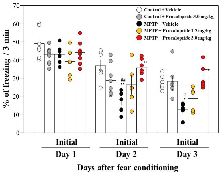
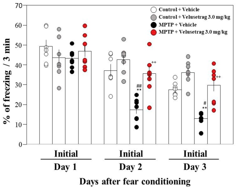
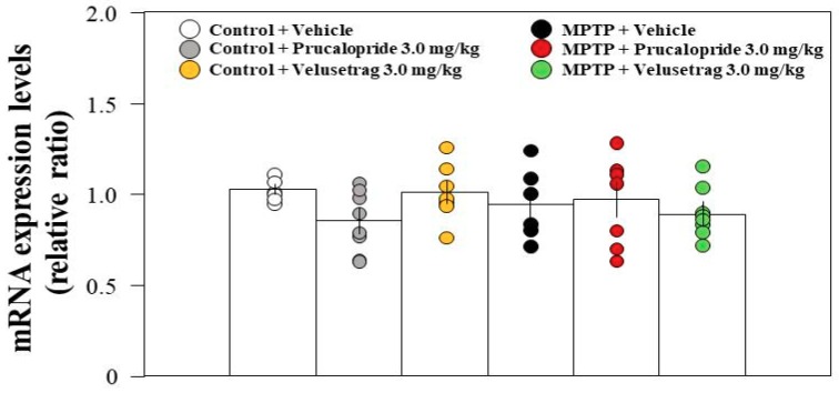
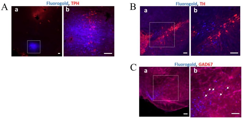
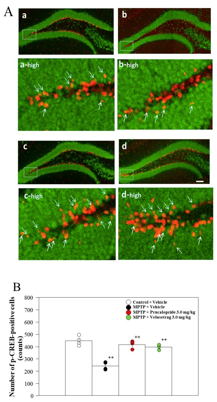
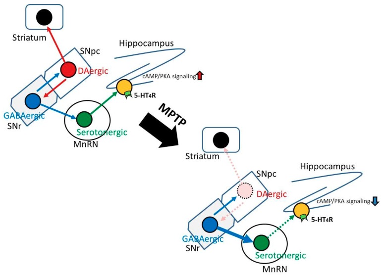
Previously, we found that 1-methyl-4-phenyl-1,2,3,6-tetrahydropyridine (MPTP)-induced Parkinson's disease (PD) model mice (PD mice) showed facilitation of hippocampal memory extinction via reduced cyclic adenosine monophosphate (cAMP)/cAMP-dependent response element-binding protein (CREB) signaling, which may cause cognitive impairment in PD. Serotonergic neurons in the median raphe nucleus (MnRN) project to the hippocampus, and functional abnormalities have been reported. In the present study, we investigated the effects of the serotonin 5-HT4 receptor (5-HT4R) agonists prucalopride and velusetrag on the facilitation of memory extinction observed in PD mice. Both 5-HT4R agonists restored facilitation of contextual fear extinction in PD mice by stimulating the cAMP/CREB pathway in the dentate gyrus of the hippocampus. A retrograde fluorogold-tracer study showed that γ-aminobutyric acid-ergic (GABAergic) neurons in the reticular part of the substantia nigra (SNr), but not dopaminergic (DAergic) neurons in the substantia nigra pars compacta (SNpc), projected to serotonergic neurons in the MnRN, which are known to project their nerve terminals to the hippocampus. It is possible that the degeneration of the SNpc DAergic neurons in PD mice affects the SNr GABAergic neurons, and thereafter, the serotonergic neurons in the MnRN, resulting in hippocampal dysfunction. These findings suggest that 5HT4R agonists could be potentially useful as therapeutic drugs for treating cognitive deficits in PD.

Keywords: 5-HT4 receptor agonist; MPTP; Parkinson’s disease; dopamine; hippocampus; median raphe nucleus (MnRN); memory extinction; reticular part of the substantia nigra (SNr); substantia nigra pars compacta (SNpc).


Conflict of interest statement

The authors declare no conflict of interest.

Figures

Figure 1
Figure 1
Effects of prucalopride on contextual fear extinction in mice. Mice were given an intraperitoneal injection of prucalopride at a dose of 1.5 or 3.0 mg/kg in saline containing 1% dimethyl sulfoxide (DMSO) or the vehicle only 2 h prior to both extinction training sessions. Data are expressed as the mean ± SEM; n = 7–8. * p < 0.05 and ** p < 0.01 vs. control + vehicle on each day, ++ p < 0.01 vs. MPTP + vehicle on each day, # p < 0.05 and ## p < 0.01 vs. control + prucalopride on each day (one-way analysis of variance (ANOVA) followed by Tukey’s post-hoc test). MPTP: 1-methyl-4-phenyl-1,2,3,6-tetrahydropyridine.
Figure 2
Figure 2
Effects of velusetrag on contextual fear extinction in mice. Mice were given an intraperitoneal injection of velusetrag at a single dose of 3.0 mg/kg in saline containing 1% DMSO or the vehicle only 2 h prior to both extinction training sessions. The data of the control + vehicle and MPTP + vehicle are the same data shown in Figure 1. Data are expressed as the mean ± SEM; n = 7–8. ** p < 0.01 vs. control + vehicle on each day, ++ p < 0.01 vs. MPTP + vehicle on each day, # p < 0.05 and ## p < 0.01 vs. control + velusetrag on each day (one-way ANOVA followed by Tukey’s post-hoc test).
Figure 3
Figure 3
RT-qPCR analysis of the hippocampal mRNA expression levels of 5-HT4 receptor (5-HT4R) in vehicle-administered control and PD mice, and in prucalopride or velusetrag-administered control and Parkinson´s disease (PD) mice. Data are expressed as the mean ± SEM: n = 6–8 per group. No significant differences were observed between groups (one-way ANOVA followed by Tukey’s post-hoc test).
Figure 4
Figure 4
Retrograde labeling of neurons following fluorogold (FG) injection into the median raphe nucleus (MnRN). (A) The square on the confocal laser-scanning microscope images under low (a) and high (b) magnification indicates the MnRN region at 3 days after FG injection. The image shows that FG (blue) was precisely injected into the MnRN. Tryptophan hydroxylase (TPH)-positive cells (red) were observed in the MnRN. Scale bar: 100 µm. (B) and (C) The square on the photomicrograph taken under low (a) and high (b) magnification indicates the substantia nigra pars compacta (SNpc) (B) and the reticular part of the reticular part of the substantia nigra (SNr) (C), which were analyzed using a confocal laser-scanning microscope at 3 days after FG injection. Scale bar: 100 µm. FG-labeled cells (blue) (B-a and -b, C-a and -b) and GAD67-positive cells (red) (C-a and -b) were co-localized in the SNr regions (indicated by white arrows) (C-b), but TH-positive cells (red) (B-a and -b) were not co-localized in the SNpc regions.
Figure 5
Figure 5
Administration of the 5-HT4R agonists to PD mice restored the decrease in cyclic adenosine monophosphate (cAMP) levels in the hippocampus. Mice were given an intraperitoneal injection of prucalopride or velusetrag at a single dose of 3.0 mg/kg in saline containing 1% DMSO or the vehicle only 2 h prior to both extinction training sessions. The hippocampus was collected immediately after the second extinction training. cAMP levels are shown as pmol/mg of trichloroacetic acid (TCA)-precipitated tissue. Data are expressed as the mean ± SEM; n = 7–8. * p < 0.05 and ** p < 0.01 vs. control + vehicle, + p < 0.05 and ++ p < 0.01 vs. MPTP + vehicle (one-way ANOVA followed by Tukey’s post-hoc test).
Figure 6
Figure 6
Analysis of the number of p-CREB-positive cells in the dentate gyrus (DG) of the hippocampus after extinction training. (A) Immunohistochemistry for p-CREB (red) and NeuN (green) in the DG after fear extinction. (a) Control + vehicle, (b) MPTP + vehicle, (c) MPTP + prucalopride 3.0 mg/kg, (d) MPTP + velusetrag 3.0 mg/kg. Scale bar = 100 µm. The images of “a-high”, “b-high”, “c-high”, and “d-high” were partially expanded from the images of the white dotted box area in a, b, c, and d, respectively. Some p-CREB-positive cells co-localize with NeuN (yellowish red, indicated by white arrows). (B) The number of p-CREB-positive cells in the DG. The mean was calculated by averaging the number of p-CREB-positive cells counted from three 50-µm sections (obtained from a position 1.82–2.02 mm posterior to the bregma). Data are expressed as the mean ± SEM; n = 3–4 per group. ** p < 0.01 vs. control + vehicle, ++ p < 0.01 vs. MPTP + vehicle (one-way ANOVA followed by Tukey’s post-hoc test).
Figure 7
Figure 7
The proposed model for cognitive impairment in PD mice and the predicted neural circuit responsible for it. The degeneration of SNpc dopaminergic (DAergic) neurons might affect γ-aminobutyric acid-ergic (GABAergic) neurons in the SNr close to the SNpc, which in turn, may influence serotonergic neurons in the MnRN receiving GABAergic neurons from the SNr, resulting in hippocampal dysfunction.
Figure 8
Figure 8
A schematic figure explaining the planning of the experiments.

References

    1. Rodriguez-Oroz M.C., Jahanshahi M., Krack P., Litvan I., Macias R., Bezard E., Obeso J.A. Initial clinical manifestations of Parkinson’s disease: Features and pathophysiological mechanisms. Lancet Neurol. 2009;8:1128–1139. doi: 10.1016/S1474-4422(09)70293-5. - DOI - PubMed
    1. Chaudhuri K.R., Healy D.G., Schapira A.H. Non-motor symptoms of Parkinson’s disease: Diagnosis and management. Lancet Neurol. 2006;5:235–245. doi: 10.1016/S1474-4422(06)70373-8. - DOI - PubMed
    1. Hou J.G., Lai E.C. Non-motor symptoms of Parkinson’s disease. Int. J. Gerontol. 2007;1:53–64. doi: 10.1016/S1873-9598(08)70024-3. - DOI
    1. Emre M. Dementia associated with Parkinson’s disease. Lancet Neurol. 2003;2:229–237. doi: 10.1016/S1474-4422(03)00351-X. - DOI - PubMed
    1. Kinoshita K., Tada Y., Muroi Y., Unno T., Ishii T. Selective loss of dopaminergic neurons in the substantia nigra pars compacta after systemic administration of MPTP facilitates extinction learning. Life Sci. 2015;137:28–36. doi: 10.1016/j.lfs.2015.07.017. - DOI - PubMed

MeSH terms