Review
. 2019 Nov 11;20(22):5639.
doi: 10.3390/ijms20225639.

Maternal Platelets—Friend or Foe of the Human Placenta?

Affiliations
Review

Maternal Platelets—Friend or Foe of the Human Placenta?

Gerit Moser et al. Int J Mol Sci. .

Abstract

Human pregnancy relies on hemochorial placentation, including implantation of the blastocyst and deep invasion of fetal trophoblast cells into maternal uterine blood vessels, enabling direct contact of maternal blood with placental villi. Hemochorial placentation requires fast and reliable hemostasis to guarantee survival of the mother, but also for the neonates. During human pregnancy, maternal platelet count decreases gradually from first, to second, and third trimester. In addition to hemodilution, accelerated platelet sequestration and consumption in the placental circulation may contribute to a decline of platelet count throughout gestation. Local stasis, turbulences, or damage of the syncytiotrophoblast layer can activate maternal platelets within the placental intervillous space and result in formation of fibrin-type fibrinoid. Perivillous fibrinoid is a regular constituent of the normal placenta which is considered to be an important regulator of intervillous hemodynamics, as well as having a role in shaping the developing villous trees. However, exaggerated activation of platelets at the maternal-fetal interface can provoke inflammasome activation in the placental trophoblast, and enhance formation of circulating platelet-monocyte aggregates, resulting in sterile inflammation of the placenta and a systemic inflammatory response in the mother. Hence, the degree of activation determines whether maternal platelets are a friend or foe of the human placenta. Exaggerated activation of maternal platelets can either directly cause or propagate the disease process in placenta-associated pregnancy pathologies, such as preeclampsia.

Keywords: placenta; platelets; preeclampsia; pregnancy.


Conflict of interest statement

The authors declare no competing interests.

Figures

Figure 1
Figure 1
Development of placental villi (a) Section through a primary villus which shows a core of cytotrophoblasts covered by the syncytiotrophoblast; (b) section through a secondary villus, which shows a core of mesenchyme covered by a two-layered trophoblast epithelium, consisting of an inner, mononucleated cytotrophoblast layer, and the outer syncytiotrophoblast; and (c) section through a tertiary villus which shows blood capillaries in the mesenchyme.
Figure 2
Figure 2
Potential route of maternal platelets into the early intervillous space. During early placenta development, maternal blood flow in uterine spiral arteries is obstructed by plugs of invaded extravillous trophoblasts (EVTs), however, maternal blood plasma and platelets can pass through narrow intertrophoblastic gaps, whereas erythrocytes and leukocytes are refrained from the intervillous space.
Figure 3
Figure 3
Localization of platelets in human first-trimester placenta in utero. An archival first-trimester human placenta in utero was obtained from hysterectomy and stained for platelet marker CD42b, as previously described by [24]. (a) The overview of the specimen shows the uterine wall composed of the perimetrium, myometrium, and endometrium, which after implantation is referred to as decidua basalis (invaded decidua) and decidua parietalis (noninvaded decidua). The chorion frondosum comprise developing placental villi, whereas the chorion laeve (smooth chorion) is the result of villous degeneration and obliteration of the intervillous space. (b) Maternal platelets can be detected on the syncytiotrophoblast layer of a villus (arrowhead) adjacent to the chorionic plate (asterisk). (c and d) An accumulation of platelets (arrowheads) can be detected in anchoring parts of trophoblast cell columns, which attach anchoring villi to the basal plate. Scale bars in b to d represent 400 µm.
Figure 4
Figure 4
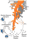
Activation of maternal platelets contributes to a proinflammatory environment in human placenta. Maternal platelets are activated by their passage through the intervillous space by local stasis, turbulences, or damage of the syncytiotrophoblast, moreover, platelets are activated by syncytiotrophoblast-derived extracellular vesicles (EVs) which are shed from the syncytiotrophoblast layer in response to oxidative and inflammatory stress. Activated platelets release ATP and provoke inflammasome activation in placental villi, leading to the release of proinflammatory cytokines into the intervillous space. Adhesion of activated platelets to the villous surface contributes to deposition of perivillous fibrin-type fibrinoids, which has been suggested as an important regulator of intervillous hemodynamics, and in shaping the microanatomy of villous trees. As soon as utero-placental blood flow is fully established, formation of platelet-monocyte aggregates induces monocytes to release proinflammatory cytokines and the antiangiogenic soluble fms-like tyrosine kinase (sFlt-1).

References

    1. Benirschke K., Burton G.J., Baergen R.N. Pathology of the Human Placenta. 6th ed. Springer-Verlag; Berlin/Heidelberg, Germany: 2012.
    1. Jauniaux E., Watson A.L., Hempstock J., Bao Y.P., Skepper J.N., Burton G.J. Onset of Maternal Arterial Blood Flow and Placental Oxidative Stress. A Possible Factor in Human Early Pregnancy Failure. Am. J. Pathol. 2000;157:2111–2122. doi: 10.1016/S0002-9440(10)64849-3. - DOI - PMC - PubMed
    1. Jauniaux E., Watson A., Ozturk O., Quick D., Burton G. In-Vivo Measurement of Intrauterine Gases and Acid-Base Values Early in Human Pregnancy. Hum. Reprod. 1999;14:2901–2904. doi: 10.1093/humrep/14.11.2901. - DOI - PubMed
    1. Burton G.J., Woods A.W., Jauniaux E., Kingdom J.C. Rheological and Physiological Consequences of Conversion of the Maternal Spiral Arteries for Uteroplacental Blood Flow during Human Pregnancy. Placenta. 2009;30:473–482. doi: 10.1016/j.placenta.2009.02.009. - DOI - PMC - PubMed
    1. Harris L.K. Review: Trophoblast-Vascular Cell Interactions in Early Pregnancy: How to Remodel a Vessel. Placenta. 2010;31:S93–S98. doi: 10.1016/j.placenta.2009.12.012. - DOI - PubMed

LinkOut - more resources