

Structure-Function Relationships of the Repeat Domains of RTX Toxins
- PMID: 31718085
- PMCID: PMC6891781
- DOI: 10.3390/toxins11110657
Structure-Function Relationships of the Repeat Domains of RTX Toxins
Abstract
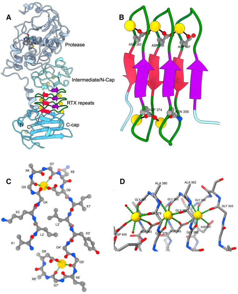
RTX proteins are a large family of polypeptides of mainly Gram-negative origin that are secreted into the extracellular medium by a type I secretion system featuring a non-cleavable C-terminal secretion signal, which is preceded by a variable number of nine-residue tandem repeats. The three-dimensional structure forms a parallel β-roll, where β-strands of two parallel sheets are connected by calcium-binding linkers in such a way that a right-handed spiral is built. The Ca2+ ions are an integral part of the structure, which cannot form without them. The structural determinants of this unique architecture will be reviewed with its conservations and variations together with the implication for secretion and folding of these proteins. The general purpose of the RTX domains appears to act as an internal chaperone that keeps the polypeptide unfolded in the calcium-deprived cytosol and triggers folding in the calcium-rich extracellular medium. A rather recent addition to the structural biology of the RTX toxin is a variant occurring in a large RTX adhesin, where this non-canonical β-roll binds to ice and diatoms.
Keywords: RTX toxin; calcium; internal chaperone; protein folding; tertiary structure; type I secretion.
Conflict of interest statement
The authors declare no conflict of interest.
Figures
References
-
- Linhartova I., Osicka R., Bumba L., Masin J., Sebo P. In: Microbial Toxins. Gopalakrishnakone P., Stiles B., Alape-Girón A., Dubreuil J.D., Mandal M., editors. Springer; Dordrecht, The Netherlands: 2015. pp. 1–29.
-
- Welch R.A. RTX Toxin Structure and Function: A Story of Numerous Anomalies and Few Analogies in Toxin Biology. Curr. Top. Microbiol. Immunol. 2001;257:85–111. - PubMed
-
- Linhartová I., Bumba L., Mašín J., Basler M., Osička R., Kamanová J., Procházková K., Adkins I., Hejnová-Holubová J., Sadílková L., et al. RTX proteins: A highly diverse family secreted by a common mechanism. FEMS Microbiol. Rev. 2010;34:1076–1112. doi: 10.1111/j.1574-6976.2010.00231.x. - DOI - PMC - PubMed
Publication types
MeSH terms
Substances
Grants and funding
LinkOut - more resources
Full Text Sources
Miscellaneous
