. 2019 Nov 12;20(1):105.
doi: 10.1186/s10194-019-1055-3.

C-fibers may modulate adjacent Aδ-fibers through axon-axon CGRP signaling at nodes of Ranvier in the trigeminal system

Affiliations

C-fibers may modulate adjacent Aδ-fibers through axon-axon CGRP signaling at nodes of Ranvier in the trigeminal system

Jacob C A Edvinsson et al. J Headache Pain. .

Abstract

Background: Monoclonal antibodies (mAbs) towards CGRP or the CGRP receptor show good prophylactic antimigraine efficacy. However, their site of action is still elusive. Due to lack of passage of mAbs across the blood-brain barrier the trigeminal system has been suggested a possible site of action because it lacks blood-brain barrier and hence is available to circulating molecules. The trigeminal ganglion (TG) harbors two types of neurons; half of which store CGRP and the rest that express CGRP receptor elements (CLR/RAMP1).

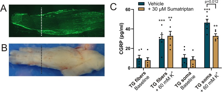
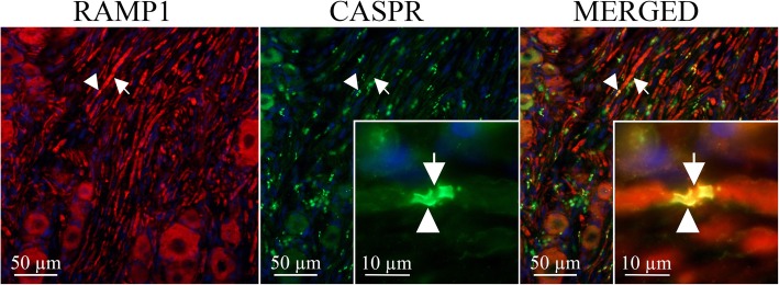
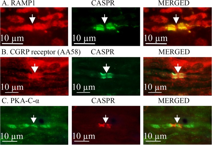
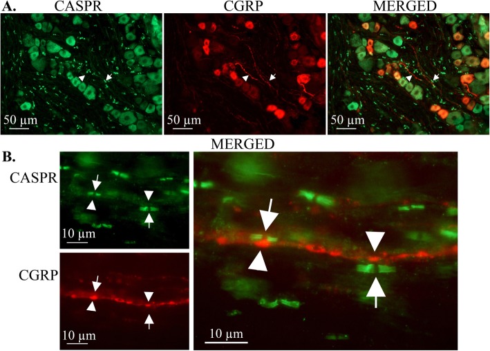
Methods: With specific immunohistochemistry methods, we demonstrated the localization of CGRP, CLR, RAMP1, and their locations related to expression of the paranodal marker contactin-associated protein 1 (CASPR). Furthermore, we studied functional CGRP release separately from the neuron soma and the part with only nerve fibers of the trigeminal ganglion, using an enzyme-linked immunosorbent assay.

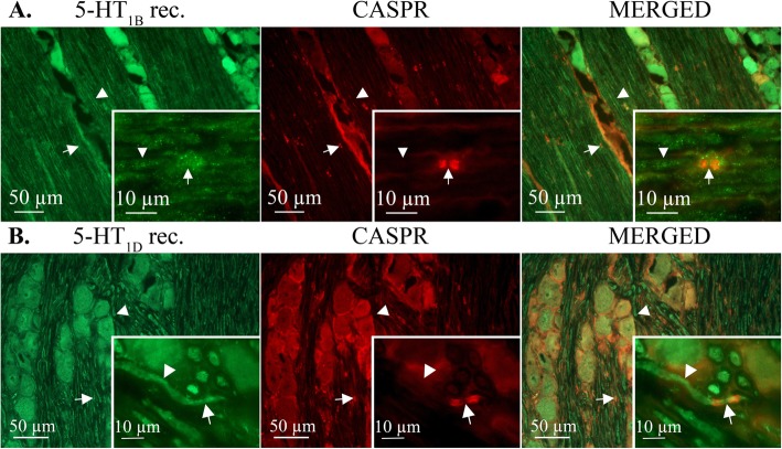
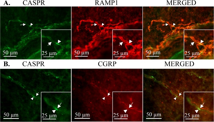
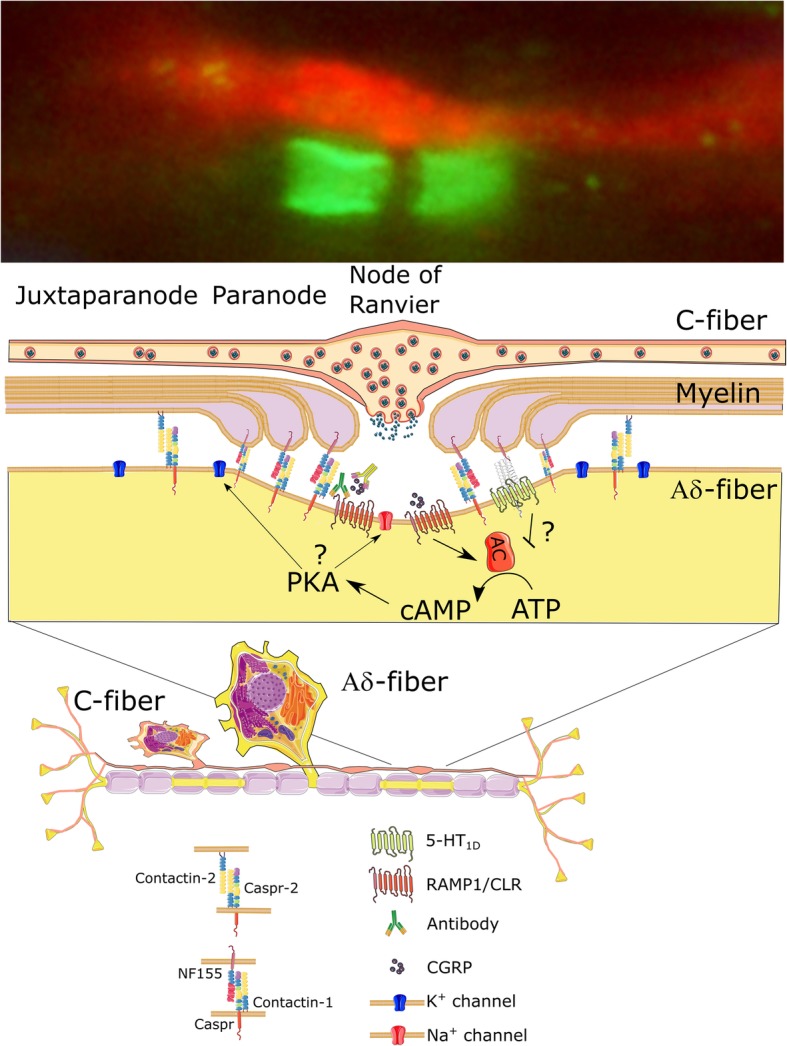
Results: Antibodies towards CGRP and CLR/RAMP1 bind to two different populations of neurons in the TG and are found in the C- and the myelinated Aδ-fibers, respectively, within the dura mater and in trigeminal ganglion (TG). CASPR staining revealed paranodal areas of the different myelinated fibers inhabiting the TG and dura mater. Double immunostaining with CASPR and RAMP1 or the functional CGRP receptor antibody (AA58) revealed co-localization of the two peptides in the paranodal region which suggests the presence of the CGRP-receptor. Double immunostaining with CGRP and CASPR revealed that thin C-fibers have CGRP-positive boutons which often localize in close proximity to the nodal areas of the CGRP-receptor positive Aδ-fibers. These boutons are pearl-like synaptic structures, and we show CGRP release from fibers dissociated from their neuronal bodies. In addition, we found that adjacent to the CGRP receptor localization in the node of Ranvier there was PKA immunoreactivity (kinase stimulated by cAMP), providing structural possibility to modify conduction activity within the Aδ-fibers.

Conclusion: We observed a close relationship between the CGRP containing C-fibers and the Aδ-fibers containing the CGRP-receptor elements, suggesting a point of axon-axon interaction for the released CGRP and a site of action for gepants and the novel mAbs to alleviate migraine.

Keywords: Aδ-fibers; C-fibers; CASPR; CGRP; Node of Ranvier,; Trigeminal ganglion.


Conflict of interest statement

The authors declare no potential conflicts of interest with respect to the research, authorship, and/or publication of this article.

Figures

Fig. 1
Fig. 1
CGRP release in vitro from two different regions isolated from the TG. The soma are located in the center of the TG (a). b The TG was cut into one area that which is fiber-rich and devoid of neuronal cell bodies (“TG fibers”) and the other that was rich in neuronal somas (“TG soma”). CGRP was increased significantly from baseline in all samples stimulated with 60 mM K+ (** p > 0.01, *** p < 0.001, paired student’s T-test). Sumatriptan significantly inhibited the CGRP release only from the from TG containing soma. Data is expressed as mean ± SEM, n = 6 for vehicle, n = 5 for sumatriptan
Fig. 2
Fig. 2
CGRP receptor localization at the paranodal region in the TGImmunohistochemical localization of the CGRP receptor protein RAMP1 (red, arrow) in myelinated axons of the trigeminal ganglion labeled with CASPR (green, arrowhead), a marker for nodes of Ranvier. Merged images reveal co-localization (yellow) of RAMP1 with CASPR at the nodes
Fig. 3
Fig. 3
Localization of CGRP receptor and PKA at the nodes of Ranvier in trigeminal axons. CGRP receptors were labeled using a RAMP1 antibody (a) or the AA58 antibody targeted to the functional CGRP receptor complex (b). Both antibodies showed co-localization with CASPR. Evidence of relevant signaling pathways is indicated by positive staining for the catalytic subunit of Protein Kinase A (PKA) at the nodes (c). Arrows indicate the node of Ranvier
Fig. 4
Fig. 4
Immunolocalization of CGRP in rat trigeminal ganglia in relation to CASPR-positive axonal nodes. a The CGRP Ab labeled smaller neurons and thin fibers (arrowhead) within the ganglia. Double staining with the CASPR Ab (arrow) showed no co-localization (merged). b Higher magnification shows a CGRP-labeled axon in close proximity to CASPR-positive axons. CGRP-containing axonal varicosities (arrow), which are putative release sites, align directly across from CASPR-positive nodes of Ranvier (arrowhead) on the adjacent axon
Fig. 5
Fig. 5
Immunolocalization of 5-HT1B and 5-HT1D receptors in the TG. Green positive immunofluorescence for 5-HT1B (a) and 5-HT1D (b) receptors throughout the Aδ-fiber, also at the node of Ranvier (monoclonal CASPR, red, arrow) No clear co-localization with CASPR was detected. Arrowheads point out immunopositive axons for both receptors
Fig. 6
Fig. 6
Expression of RAMP1 and CGRP in relation to the node of Ranvier in rat dura mater spreads. RAMP1 (a) and CGRP (b) had similar localization at the node of Ranvier in the rat dura mater as for the TG. Top; CASPR (green, arrowhead), RAMP1 (red, arrow). Bottom; CASPR (green, arrowhead), CGRP (red, arrow)
Fig. 7
Fig. 7
Schematic overview of the possible modulation by C-fibers on adjacent Aδ-fibers through axon-axon signaling at nodes of Ranvier in the trigeminal system. CGRP is released from the C-fiber bouton and diffuses into the node of Ranvier. Activation of the CGRP receptor causes increase in cyclic AMP, which could alter the conductivity of Na+ or K+ channels through Phosphokinase A (PKA). Furthermore, there is a possibility that 5-HT receptors could negatively modulate the cyclic AMP at the node. Alterations in Na+ or K+ channels could modulate the nodal threshold of activation. The schematic also includes a hypothesis on the mode of action of the anti-migraine antibody treatments

References

    1. Edvinsson L, Haanes KA, Warfvinge K, Krause DN. CGRP as the target of new migraine therapies - successful translation from bench to clinic. Nat Rev Neurol. 2018;14(6):338–350. doi: 10.1038/s41582-018-0003-1. - DOI - PubMed
    1. Edvinsson L. The Trigeminovascular pathway: role of CGRP and CGRP receptors in migraine. Headache. 2017;57(Suppl 2):47–55. doi: 10.1111/head.13081. - DOI - PubMed
    1. Olesen J, Diener HC, Husstedt IW, Goadsby PJ, Hall D, Meier U, et al. Calcitonin gene-related peptide receptor antagonist BIBN 4096 BS for the acute treatment of migraine. N Engl J Med. 2004;350(11):1104–1110. doi: 10.1056/NEJMoa030505. - DOI - PubMed
    1. Lattanzi S, Brigo F, Trinka E, Vernieri F, Corradetti T, Dobran M, et al. Erenumab for preventive treatment of migraine: a systematic review and meta-analysis of efficacy and safety. Drugs. 2019;79(4):417–431. doi: 10.1007/s40265-019-01069-1. - DOI - PubMed
    1. Boado RJ, Zhou QH, Lu JZ, Hui EK, Pardridge WM. Pharmacokinetics and brain uptake of a genetically engineered bifunctional fusion antibody targeting the mouse transferrin receptor. Mol Pharm. 2010;7(1):237–244. doi: 10.1021/mp900235k. - DOI - PMC - PubMed

MeSH terms

Substances

Grants and funding