

Spectrum of clinical and radiographic findings in patients with diagnosis of H1N1 and correlation with clinical severity
- PMID: 31718571
- PMCID: PMC6852716
- DOI: 10.1186/s12879-019-4592-0
Spectrum of clinical and radiographic findings in patients with diagnosis of H1N1 and correlation with clinical severity
Abstract
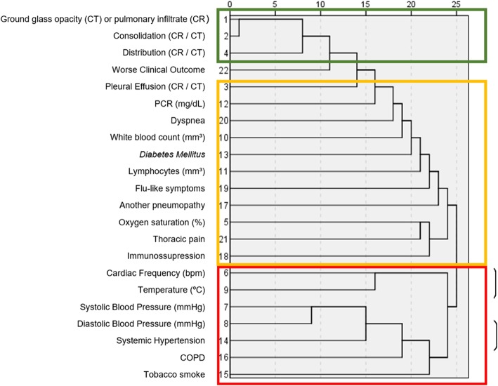
Background: The aim of this study was to evaluate the correlation between clinical and imaging findings with a worse clinical outcome in patients with a confirmed diagnosis of H1N1 influenza A virus.
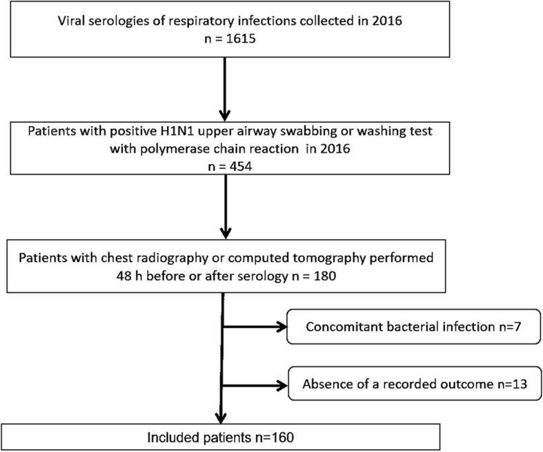
Methods: Patients with a positive viral test for influenza A H1N1 in 2016 and chest radiography (CR) and/or computed tomography (CT) results had clinical and imaging data reviewed. Hospitalization, admission to the intensive care unit or death were defined as worse clinical outcomes. The association between clinical and imaging features and the worse outcome was calculated in a logistical regression model.
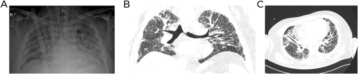
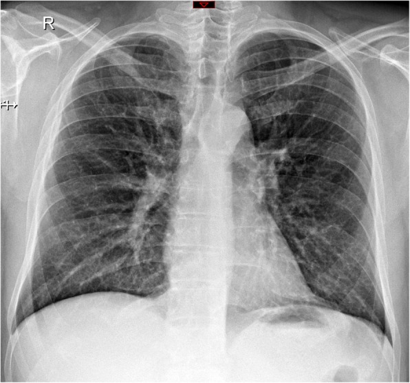
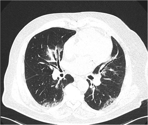
Results: Eighty of 160 (50%) patients were men, with a mean age of 43 ± 19 years. The most common symptoms were as follows: flu-like symptoms 141/160 (88%), dyspnea (25/160, 17%), and thoracic pain (7/160, 5%). Abnormalities on CR were detected in 8/110 (7%) patients, and 43/59 (73%) patients had an abnormal CT. The following variables were associated with worse clinical outcomes: the presence of diabetes mellitus (DM), hypertension, dyspnea, thoracic pain, abnormal CR or CT regardless of the type of finding, CT with consolidation or ground glass opacity.
Conclusions: The presence of DM, hypertension, dyspnea, thoracic pain, or an abnormal CR or CT on admission were associated with worse clinical outcomes in patients with H1N1 influenza A virus infection. Thus, the use of readily accessible clinical and imaging features on admission may have a role in the evaluation of patients with H1N1 infection.
Keywords: H1N1; Infection; Influenza; Outcome; Radiology.
Conflict of interest statement
The authors declare that they have no competing interests.
Figures
References
-
- Brasil registra 3978 de H1N1 em 2016. Agência Brasil [http://agenciabrasil.ebc.com.br/geral/noticia/2016-06/brasil-registra-39...]. Accessed 29 Nov 2018.
MeSH terms
LinkOut - more resources
Full Text Sources
Medical
