. 2019 Nov 12;9(1):16595.
doi: 10.1038/s41598-019-52656-x.

The gene structure and hypervariability of the complete Penaeus monodon Dscam gene

Affiliations

The gene structure and hypervariability of the complete Penaeus monodon Dscam gene

Kantamas Apitanyasai et al. Sci Rep. .

Abstract

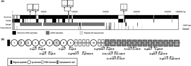
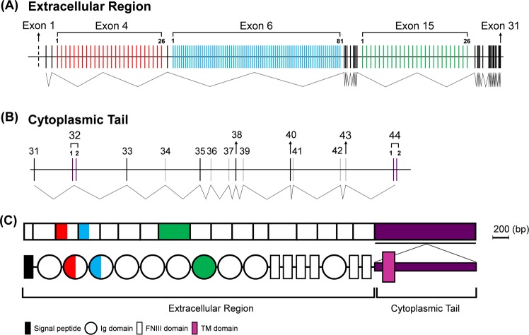
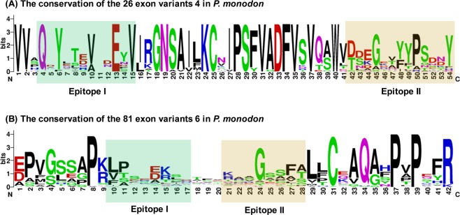
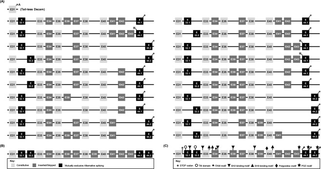
Using two advanced sequencing approaches, Illumina and PacBio, we derive the entire Dscam gene from an M2 assembly of the complete Penaeus monodon genome. The P. monodon Dscam (PmDscam) gene is ~266 kbp, with a total of 44 exons, 5 of which are subject to alternative splicing. PmDscam has a conserved architectural structure consisting of an extracellular region with hypervariable Ig domains, a transmembrane domain, and a cytoplasmic tail. We show that, contrary to a previous report, there are in fact 26, 81 and 26 alternative exons in N-terminal Ig2, N-terminal Ig3 and the entirety of Ig7, respectively. We also identified two alternatively spliced exons in the cytoplasmic tail, with transmembrane domains in exon variants 32.1 and 32.2, and stop codons in exon variants 44.1 and 44.2. This means that alternative splicing is involved in the selection of the stop codon. There are also 7 non-constitutive cytoplasmic tail exons that can either be included or skipped. Alternative splicing and the non-constitutive exons together produce more than 21 million isoform combinations from one PmDscam locus in the P. monodon gene. A public-facing database that allows BLAST searches of all 175 exons in the PmDscam gene has been established at http://pmdscam.dbbs.ncku.edu.tw/ .


Conflict of interest statement

The authors declare no competing interests.

Figures

Figure 1
Figure 1
Strategies and genomic sequencing methods. (A) Construction and characterization of the polished M2 assembly of the complete P. monodon genome that was used to produce a draft sequence of the PmDscam Dscam gene. (B) Analysis steps used to determine the PmDscam cytoplasmic tail and location of the cytoplasmic tail exons.
Figure 2
Figure 2
Schematic diagram of the Penaeus monodon Dscam gene structure. (A) PmDscam genomic DNA spans 266 kbp. PacBio and Illumina sequencing were used to characterize the entire gene, with Sanger sequencing used to fill some gaps and confirm sequences. Transcriptomics data were used to identify the cytoplasmic tail and some parts of the extracellular region. Samples used for sequencing were derived from both genomic DNA (black) and cDNA (dark grey) samples. The gap near the N-terminal corresponds to a part of the 5′-UTR that we were unable to find in the gene. The boxes above the schematic show the location of five other parts of the gene that contain unknown repeat sequences (N). (B) Location of primers for PCR amplification and Sanger sequencing of the PmDscam gene. Samples were extracted from both genomic DNA and cDNA.
Figure 3
Figure 3
Organization of the PmDscam gene. The PmDscam gene consists of 175 exons and/or exon variants: 31 exons are constitutive (black lines), 7 exons (all in the cytoplasmic tail) can be either inserted or skipped (gray lines), and 137 exon variants are subject to mutually exclusive alternative splicing (colored lines). (A) The PmDscam extracellular region is encoded by exon 1 to exon 31. The variable regions are exon 4 (red), exon 6 (blue) and exon 15 (green), which contain 26 variants, 81 variants and 26 variants, respectively. The dashed line represents exon 1, which was not identified from the P. monodon gene. (B) The cytoplasmic tail is encoded by exon 31 to exon 44. The variable regions are exon 32 and exon 44, with each of these two exons derived from two mutually exclusive variants; that is, after RNA splicing, each transcript contains one of the alternative variants for each of these exons. (C) The extracellular region of PmDscam mRNA (upper panel) contains both constitutive exons (white) and exons that are subject to mutually exclusive alternative splicing. Alternatively spliced exons encode the N-terminal half of Ig2 (red), the N-terminal half of Ig3 (blue), and the entirety of Ig7 (green). In the cytoplasmic tail (purple), both the transmembrane domain (exon 32) and exon 44 are subject to mutually exclusive alternative splicing. The PmDscam protein structure (lower panel) is comprised of the extracellular region, which contains 10 immunoglobulin (Ig) domains and six fibronectin type 3 (FNIII) domains, followed by the cytoplasmic tail.
Figure 4
Figure 4
Multiple amino acid sequence alignments of each of the PmDscam extracellular variable regions. (A) 26 variants encode the N-terminal Ig2 domain in the Ig2 exon 4 cluster. (B) 81 variants encode the N-terminal Ig3 domain in the Ig3 exon 6 cluster. (C) 26 variants encode the entire Ig7 domain in the Ig7 exon 15 cluster. The total number of amino acids for each isoform is indicated on the right. Identical (black) and similar (grey and light grey) amino acids are indicated. Exon#: the exon numbers correspond to the exon’s location in the PmDscam gene.
Figure 5
Figure 5
Differential sequence conservation of epitopes I and II of PmDscam. (A) Sequence logo representation of the conservation of exon 4 variants in P. monodon. (B) Sequence logo representation of the conservation of exon 6 variants in P. monodon. Bits on the y-axis indicate units of evolutionary conservation.
Figure 6
Figure 6
PmDscam isoform expression in hemocytes and nerve tissue. (A) Location of primers for PCR amplification and sequencing of PmDscam cDNA domain structure. (B) Exon 4 variants (C) exon 6 variants and (D) exon 15 variants detected in hemocyte (hcy) and nerve (nev) cDNA. Hemocyte and nerve samples were collected from 10 individual shrimp for total RNA extraction and cDNA synthesis. Twenty individual cDNA clones were obtained from each sample and their exon 4-exon 15 (Ig2-Ig7 domain) was sequenced. Red boxes represent detection of the isoform while green boxes represent non-detection of the isoform.
Figure 7
Figure 7
Organization of the PmDscam cytoplasmic tail. Exon numbers were determined according to the location of the exon in the PmDscam gene. (A) Schematic diagram showing the cytoplasmic tail exon combinations of 20 PmDscam contigs obtained from transcriptomics data. (B) Summary of exon types. Constitutive exons, inserted or skipped exons and alternative exon variants are shown as light grey boxes, dark grey boxes, and black boxes, respectively. (C) Cytoplasmic tail exons showing locations of common functional domains and motifs. Asterisks indicate a stop codon.
Figure 8
Figure 8
The amino acid sequence of a PmDscam isoform. In the extracellular region, putative signal peptides are in italics, and Ig domains are shaded light grey. The variable sequences in Ig2, Ig3 and Ig7 are in bold. FNIII domains are boxed. A conserved RGD motif is indicated by underlining. In the cytoplasmic tail, which is rendered in reverse contrast, the constitutive and optional domains are shaded black, while the mutually exclusive alternatively spliced domains are in bold against a light grey background. The asterisk indicates the stop codon.

References

    1. Yamakawa K, et al. DSCAM: a novel member of the immunoglobulin superfamily maps in a Down syndrome region and is involved in the development of the nervous system. Hum Mol Genet. 1998;7:227–37. doi: 10.1093/hmg/7.2.227. - DOI - PubMed
    1. Watson FL, et al. Extensive diversity of Ig-superfamily proteins in the immune system of insects. Science. 2005;309:1874–8. doi: 10.1126/science.1116887. - DOI - PubMed
    1. Dong Y, Taylor HE, Dimopoulos G. AgDscam, a hypervariable immunoglobulin domain-containing receptor of the Anopheles gambiae innate immune system. PLoS Biol. 2006;4:e229. doi: 10.1371/journal.pbio.0040229. - DOI - PMC - PubMed
    1. Armitage SA, Peuss R, Kurtz J. Dscam and pancrustacean immune memory – a review of the evidence. Dev Comp Immunol. 2015;48:315–23. doi: 10.1016/j.dci.2014.03.004. - DOI - PubMed
    1. Schmucker D, et al. Drosophila Dscam is an axon guidance receptor exhibiting extraordinary molecular diversity. Cell. 2000;101:671–84. doi: 10.1016/S0092-8674(00)80878-8. - DOI - PubMed

Publication types

Substances