. 2019 Nov 12;9(1):16574.
doi: 10.1038/s41598-019-53242-x.

Dietary short-chain fatty acid intake improves the hepatic metabolic condition via FFAR3

Affiliations

Dietary short-chain fatty acid intake improves the hepatic metabolic condition via FFAR3

Hidenori Shimizu et al. Sci Rep. .

Abstract

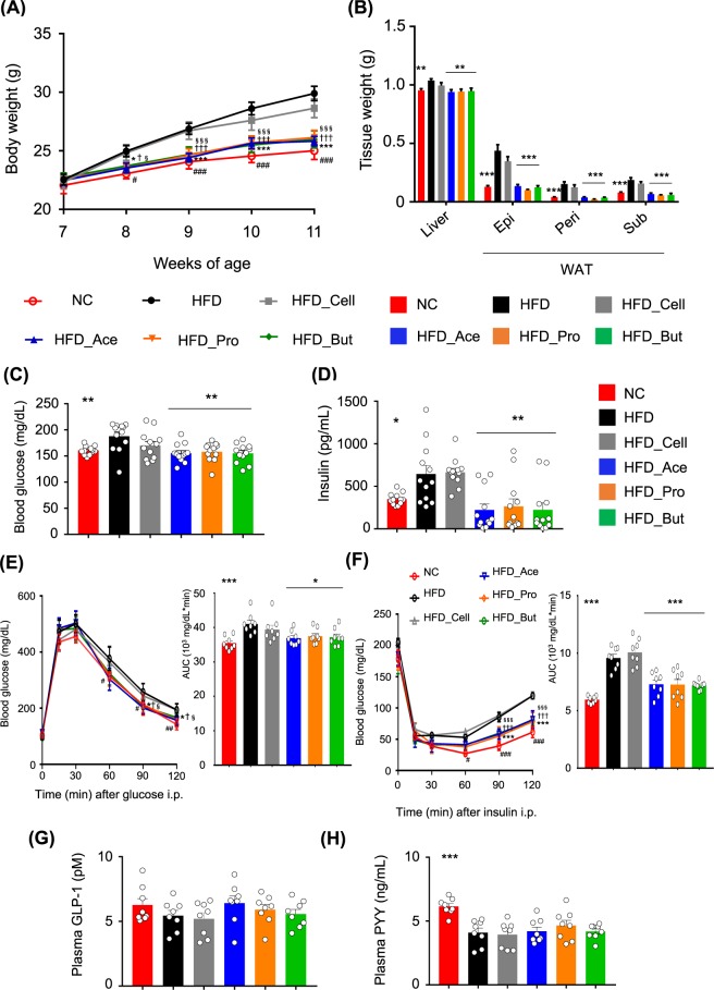
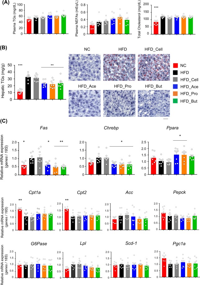
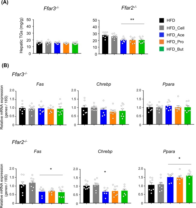
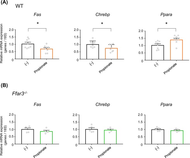
Fermented foods represent a significant portion of human diets with several beneficial effects. Foods produced by bacterial fermentation are enriched in short-chain fatty acids (SCFAs), which are functional products of dietary fibers via gut microbial fermentation. In addition to energy sources, SCFAs also act as signaling molecules via G-protein coupled receptors such as FFAR2 and FFAR3. Hence, dietary SCFAs in fermented foods may have a direct influence on metabolic functions. However, the detailed mechanism by dietary SCFAs remains unclear. Here, we show that dietary SCFAs protected against high-fat diet-induced obesity in mice in parallel with increased plasma SCFAs without changing cecal SCFA or gut microbial composition. Dietary SCFAs suppressed hepatic weight and lipid synthesis. These effects were abolished in FFAR3-deficient mice but not FFAR2-deficient. Thus, SCFAs supplementation improved hepatic metabolic functions via FFAR3 without influencing intestinal environment. These findings could help to promote the development of functional foods using SCFAs.


Conflict of interest statement

The authors declare no competing interests.

Figures

Figure 1
Figure 1
Short-chain fatty acid (SCFA) supplementation exerts metabolic benefits. Body weight changes (A), liver and white adipose tissue (WAT) weights (B), blood glucose (C), plasma insulin (D), glucose tolerance test (GTT) (E), insulin tolerance test (ITT) (F), GLP-1 (G), and PYY levels (H) measured after 4 weeks of normal chow (NC) or high-fat diet (HFD) feeding supplemented with 5% SCFAs. All data are presented as the means ± SEM (n = 8–12). Dunnett’s test; ***P < 0.001, **P < 0.01, and *P < 0.05, compared with HFD. ###P < 0.001, ##P < 0.01, and #P < 0.05 (HFD vs. NC), ***P < 0.001 and *P < 0.05 (HFD vs. Ace), †††P < 0.001 and P < 0.05 (HFD vs. Pro), §§§P < 0.001 and §P < 0.05 (HFD vs. But) (A,E,F). Epi: epididymal tissue, Peri: perirenal tissue, Sub: subcutaneous tissue.
Figure 2
Figure 2
Dietary short-chain fatty acid (SCFA) intake improves hepatic metabolic conditions. Plasma triglycerides, non-esterified fatty acids (NEFAs), and total cholesterol concentrations (A), hepatic triglyceride contents and histology of hepatocytes based on hematoxylin-eosin (H&E) oil red O stanning. Scale bar, 50 μm (B), and mRNA expression levels of hepatic energy metabolism-related genes (C) measured after 4 weeks of normal chow (NC) or high-fat diet (HFD) feeding supplemented with 5% SCFAs. All data are presented as the means ± SEM (n = 8). Dunnett’s test; ***P < 0.001, **P < 0.01, and *P < 0.05, compared with HFD.
Figure 3
Figure 3
FFAR3 deficiency abolishes dietary short-chain fatty acid (SCFA) intake-induced metabolic benefits. Body weight changes (A), liver and white adipose tissue (WAT) weights (B), blood glucose (C), and plasma insulin (D) levels in Ffar3−/− (n = 10) and Ffar2−/− (n = 8–10) mice measured after 4 weeks of high-fat diet (HFD) feeding supplemented with 5% SCFAs. All data are presented as the means ± SEM. Dunnett’s test; ***P < 0.001, **P < 0.01, and *P < 0.05, compared with HFD. **P < 0.01 (HFD vs. Ace), ††P < 0.01 (HFD vs. Pro), §§P < 0.01 (HFD vs. But) (A). Epi: epididymal tissue, Peri: perirenal tissue, Sub: subcutaneous tissue.
Figure 4
Figure 4
FFAR3 deficiency abolishes dietary short-chain fatty acid (SCFA) intake-induced expression change of hepatic lipid metabolism-related genes. Hepatic triglycerides contents (A) and mRNA expression levels of hepatic lipid metabolism-related genes (B) in Ffar3−/− and Ffar2−/− mice (n = 7–8) after 4 weeks of high-fat diet (HFD) feeding supplemented with 5% SCFAs. All data are presented as the means ± SEM. Dunnett’s test; **P < 0.01 and *P < 0.05, compared with HFD.
Figure 5
Figure 5
FFAR3 deficiency abolishes acute short-chain fatty acid (SCFA) injection-induced expression changes in hepatic lipid metabolism-related genes. mRNA expression levels of hepatic lipid metabolism-related genes in wild-type (WT) (n = 8) (A) and Ffar3−/− (B) mice (n = 5) at 24 h after intraperitoneal phosphate buffered saline (for control group) or propionate administration (1 g/kg body weight) under high-fat diet (HFD) feeding. All data are presented as the means ± SEM. Student’s t-test; *P < 0.05, compared with (−).

Similar articles

Cited by

References

    1. Li X, Guo J, Ji K, Zhang P. Bamboo shoot fiber prevents obesity in mice by modulating the gut microbiota. Sci. Rep. 2016;6:32953. doi: 10.1038/srep32953. - DOI - PMC - PubMed
    1. Miyamoto J, et al. Barley beta-glucan improves metabolic condition via short-chain fatty acids produced by gut microbial fermentation in high fat diet fed mice. PLoS One. 2018;13:e0196579. doi: 10.1371/journal.pone.0196579. - DOI - PMC - PubMed
    1. Chambers ES, Preston T, Frost G, Morrison DJ. Role of gut microbiota-generated short-chain fatty acids in metabolic and cardiovascular health. Curr. Nutr. Rep. 2018;7:198–206. doi: 10.1007/s13668-018-0248-8. - DOI - PMC - PubMed
    1. Fernandes J, Su W, Rahat-Rozenbloom S, Wolever TM, Comelli EM. Adiposity, gut microbiota and faecal short chain fatty acids are linked in adult humans. Nutr. Diabetes. 2014;4:e121. doi: 10.1038/nutd.2014.23. - DOI - PMC - PubMed
    1. Wolfe BE, Dutton RJ. Fermented foods as experimentally tractable microbial ecosystems. Cell. 2015;161:49–55. doi: 10.1016/j.cell.2015.02.034. - DOI - PubMed

Publication types

LinkOut - more resources