. 2021 Aug;26(8):4383-4393.
doi: 10.1038/s41380-019-0586-y. Epub 2019 Nov 12.

Dissociable neural substrates of opioid and cocaine use identified via connectome-based modelling

Affiliations

Dissociable neural substrates of opioid and cocaine use identified via connectome-based modelling

Sarah D Lichenstein et al. Mol Psychiatry. 2021 Aug.

Abstract

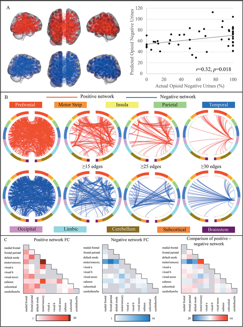
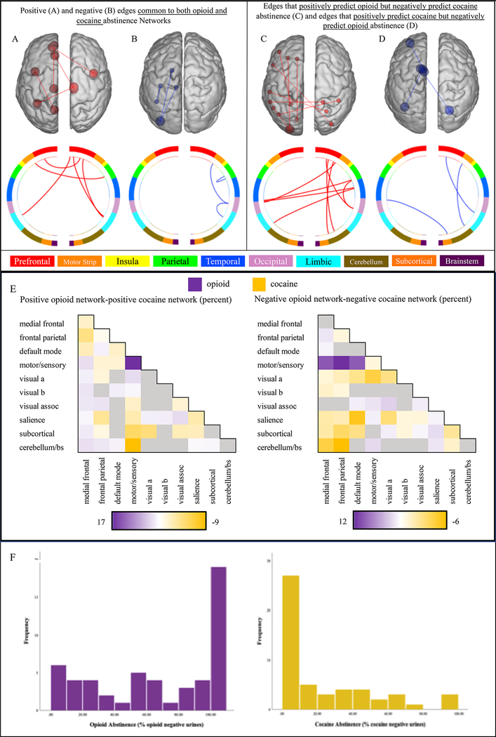
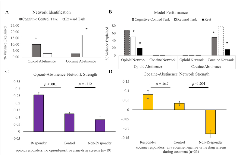
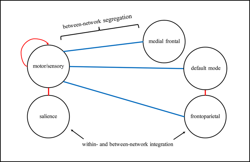
Opioid use disorder is a major public health crisis. While effective treatments are available, outcomes vary widely across individuals and relapse rates remain high. Understanding neural mechanisms of treatment response may facilitate the development of personalized and/or novel treatment approaches. Methadone-maintained, polysubstance-using individuals (n = 53) participated in fMRI scanning before and after substance-use treatment. Connectome-based predictive modeling (CPM)-a recently developed, whole-brain approach-was used to identify pretreatment connections associated with abstinence during the 3-month treatment. Follow-up analyses were conducted to determine the specificity of the identified opioid abstinence network across different brain states (cognitive vs. reward task vs. resting-state) and different substance use outcomes (opioid vs. cocaine abstinence). Posttreatment fMRI data were used to assess network changes over time and within-subject replication. To determine further clinical relevance, opioid abstinence network strength was compared with healthy subjects (n = 38). CPM identified an opioid abstinence network (p = 0.018), characterized by stronger within-network motor/sensory connectivity, and reduced connectivity between the motor/sensory network and medial frontal, default mode, and frontoparietal networks. This opioid abstinence network was anatomically distinct from a previously identified cocaine abstinence network. Relationships between abstinence and opioid and cocaine abstinence networks replicated across multiple brain states but did not generalize across substances. Network connectivity measured at posttreatment related to abstinence at 6-month follow-up (p < 0.009). Healthy comparison subjects displayed intermediate network strengths relative to treatment responders and nonresponders. These data indicate dissociable anatomical substrates of opioid vs. cocaine abstinence. Results may inform the development of novel opioid-specific treatment approaches to combat the opioid epidemic.


Figures

Figure 1 –
Figure 1 –
Positive and negative opioid abstinence networks Figure 1A displays the positive opioid abstinence network (red), for which stronger connectivity predicts more opioid abstinence during treatment and the negative opioid abstinence network (blue), in which decreased connectivity is associated with more opioid abstinence. Larger spheres correspond to higher degree nodes. There was a significant association between CPM-predicted opioid abstinence (y-axis) and the observed percentage of opioid-negative urines during treatment (x-axis; p=.018). Figure 1B displays circle plots summarizing positive (red) and negative (blue) opioid abstinence network anatomy based on overlap with macroscale brain regions. Regions are organized according to their anatomical location, with more anterior regions are at the top of each plot, and more ventral and posterior regions displayed towards the bottom. The first set of plots contain all network edges, subsequent plots are thresholded to include only higher degree nodes. Figure 1C summarizes positive and negative opioid abstinence network anatomy based on overlap with canonical neural networks. Cells along the diagonal represent within-network connectivity and the remaining cells correspond to between-network connectivity. Color bars correspond to the percentage of each network accounted for by edges within/between each canonical neural network. Darker colors indicate a higher percentage of edges. The last figure shows the percentage of positive versus negative edges that correspond to different canonical neural networks. Red cells indicate relatively more positive network edges and blue cells indicate relatively more negative network edges.
Figure 2 –
Figure 2 –
Anatomical specificity of opioid and cocaine abstinence networks Figure 2A-D compares the anatomy of the opioid and cocaine abstinence networks based on their overlap with macroscale brain regions. Figure 2A depicts edges that belong to both the positive opioid and positive cocaine abstinence networks, and figure 2B shows edges that are common to both the negative opioid and the negative cocaine abstinence network. Figure 2C shows edges that positively predict opioid but negatively predict cocaine abstinence, and figure 2D displays edges that positively predict cocaine but negatively predict opioid abstinence. Figure 2E compares the anatomy of the opioid and cocaine abstinence networks based on their overlap with canonical neural networks. Color bars correspond to the difference between the percentage of the positive and negative opioid versus cocaine abstinence network accounted for by edges within and between each canonical network. Purple cells indicate relatively more edges in the opioid abstinence network and orange cells indicate relatively more edges in the cocaine abstinence network. Figure 2F displays frequency distributions of the percent of opioid and cocaine negative urines collected across the 12-week treatment period. Percent of opioid negative urines was not significantly correlated with the percent of cocaine negative urines (Spearman rho=.082, p=.565).
Figure 3 –
Figure 3 –
Brain state manipulation and comparison with healthy controls Figure 3A demonstrates that CPM analyses successfully predict opioid abstinence using cognitive control - but not reward task - data, whereas cocaine abstinence is successfully predicted by reward task – but not cognitive control task – data. Figure 3B demonstrates that both opioid and cocaine abstinence networks can predict substance use outcomes (opioid or cocaine use, respectively) across brain states, and perform better with task versus resting-state data. Associations between the strength of each network in the brain state used for network generation (opioid abstinence network strength in cognitive control task data and cocaine abstinence network strength in reward task data) and abstinence are plotted for reference using dotted lines. Figure 3C-D compares opioid (3C) and cocaine (3D) network strength (y-axis) between treatment responders, treatment non-responders, and HCs. Because patients were stably maintained on methadone at the time of study enrollment, patients were classified as opioid treatment responders if they had no opioid positive urines during treatment. In contrast, patients were actively using cocaine at the time of study enrollment, so patients were classified as cocaine treatment responders if they had any cocaine negative drug screens during treatment. Error bars indicate the standard error of the mean.
Figure 4 –
Figure 4 –
Network model of opioid abstinence network Figure 4 presents a network model summarizing the dominant connections of the opioid abstinence network. Stronger connectivity (red) within the motor/sensory network and between motor/sensory and salience, and default mode and frontoparietal networks predicted greater within-treatment opioid abstinence, and reduced connectivity (blue) between the motor/sensory and medial frontal, default mode, and frontoparietal networks predicted greater within-treatment opioid abstinence.

References

    1. McCarty D, Priest KC, Korthuis PT. Treatment and Prevention of Opioid Use Disorder: Challenges and Opportunities. Annu Rev Public Health 2018; 39: 525–541. - PMC - PubMed
    1. McCabe SE, West BT, Jutkiewicz EM, Boyd CJ. Multiple DSM-5 substance use disorders: A national study of US adults. Hum Psychopharmacol 2017; 32(5). - PMC - PubMed
    1. Van den Brink W, Haasen C. Evidenced-based treatment of opioid-dependent patients. Can J Psychiatry 2006; 51(10): 635–646. - PubMed
    1. Connery HS. Medication-assisted treatment of opioid use disorder: review of the evidence and future directions. Harv Rev Psychiatry 2015; 23(2): 63–75. - PubMed
    1. Moningka H, Lichenstein S, Worhunsky PD, DeVito EE, Scheinost D, Yip SW. Can neuroimaging help combat the opioid epidemic? A systematic review of clinical and pharmacological challenge fMRI studies with recommendations for future research. Neuropsychopharmacology 2019; 44(2): 259–273. - PMC - PubMed

Publication types