. 2018 May 2;11(3):163-174.
doi: 10.1007/s12195-018-0526-y. eCollection 2018 Jun.

Mouse Keratinocytes Without Keratin Intermediate Filaments Demonstrate Substrate Stiffness Dependent Behaviors

Affiliations

Mouse Keratinocytes Without Keratin Intermediate Filaments Demonstrate Substrate Stiffness Dependent Behaviors

Hoda Zarkoob et al. Cell Mol Bioeng. .

Abstract

Introduction: Traditionally thought to serve active vs. passive mechanical functions, respectively, a growing body of evidence suggests that actin microfilament and keratin intermediate filament (IF) networks, together with their associated cell-cell and cell-matrix anchoring junctions, may have a large degree of functional interdependence. Therefore, we hypothesized that the loss of keratin IFs in a knockout mouse keratinocyte model would affect the kinematics of colony formation, i.e., the spatiotemporal process by which individual cells join to form colonies and eventually a nascent epithelial sheet.

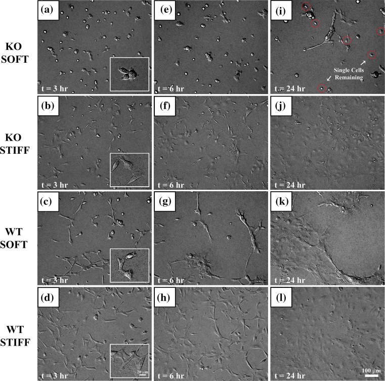
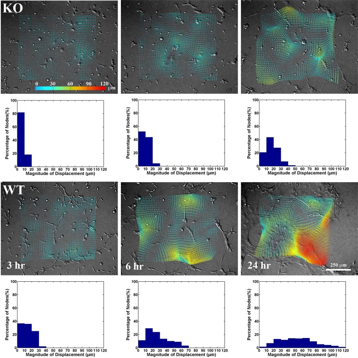
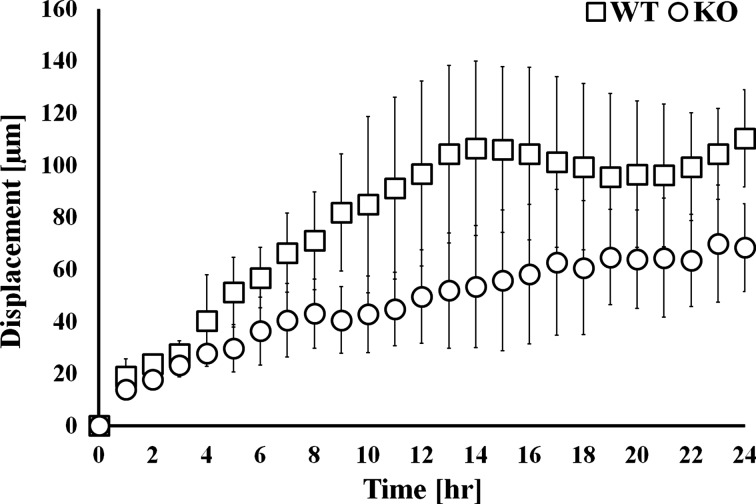
Methods: Time-lapse imaging and deformation tracking microscopy was used to observe colony formation for both wild type (WT) and keratin-deficient knockout (KO) mouse keratinocytes over 24 h. Cells were cultured under high calcium conditions on collagen-coated substrates with nominal stiffnesses of ~ 1.2 kPa (soft) and 24 kPa (stiff). Immunofluorescent staining of actin and selected adhesion proteins was also performed.

Results: The absence of keratin IFs markedly affected cell morphology, spread area, and cytoskeleton and adhesion protein organization on both soft and stiff substrates. Strikingly, an absence of keratin IFs also significantly reduced the ability of mouse keratinocytes to mechanically deform the soft substrate. Furthermore, KO cells formed colonies more efficiently on stiff vs. soft substrates, a behavior opposite to that observed for WT keratinocytes.

Conclusions: Collectively, these data are strongly supportive of the idea that an interdependence between actin microfilaments and keratin IFs does exist, while further suggesting that keratin IFs may represent an important and under-recognized component of keratinocyte mechanosensation and the force generation apparatus.

Keywords: Force; Intermediate filaments; Keratins; Mechanosensing; Polyacrylamide gels; Traction microscopy.


Figures

Figure 1
Figure 1
KO and WT cell morphology on soft and stiff substrate at t = 3, 6, and 24 h after plating the cells onto soft and stiff PA gels. KO soft (a, e, i), KO stiff (b, f, j), WT soft (c, g, k), and WT stiff (d, h, l) PA gels show distinct differences in morphology and colony forming behaviors. KO cells on soft PA gels were more rounded with fewer protrusions compared to WT and stiff gels, whereas the cells were more elongated and spindle-shaped (see insets a, b, c, d for higher magnification). Single cells (red circles) were still present on KO soft gels after 24 h (i), but not for the other three conditions (j, k, l).
Figure 2
Figure 2
Comparison of F-actin (red) organization and cell nuclei (blue) at t = 3, 6, and 24 h. Distinct differences in F-actin organization were apparent as a function of cell genotype and PA gel stiffness at t = 3, 6, and 24 h, respectively. Cortical actin could be found in KO on stiff PA gels (b, f, j) and in WT on both soft (c, g, k) and stiff (d, h, l) PA gels. In contrast, only cytoplasmic aggregates of actin were observed for KO on soft PA gels. Images (m, n, o, p) are larger fields of view of images (i, j, k, l).
Figure 3
Figure 3
Comparison of cell–ECM and cell–cell adhesions at t = 24 h. (a–d) Integrin β1 subunit (green) and vinculin (red). (e–h) Integrin β4 subunit (green) and vinculin (red). (i–l) Desmoplakin (green) and E-cadherin (red).
Figure 4
Figure 4
Displacement tracking (DTM) of KO and WT on soft PA gels at 3, 6, and 24 h. The tracked displacements are overlaid on the DIC images. Arrows show the direction and magnitude of substrate displacements. Below each image is the corresponding histogram showing the percentages of tracked substrate locations (i.e., nodes) as a function of substrate displacements. Chi-squared tests indicate that the magnitude of the displacements increases significantly with time (p < 0.001), and that displacements are significantly higher for WT than for KO (p < 0.001).
Figure 5
Figure 5
Maximum displacements produced by KO and WT on soft PA gels over time. Data are presented as the average and standard deviation of three samples for each cell genotype. A two-way ANOVA indicates that maximum displacements were significantly higher (p < 0.001) in WT compared to KO.
Figure 6
Figure 6
Percentages of single cells remaining in the field of view over time. The decrease was faster for WT compared to KO. Single KO cells still remained on soft PA gels after 24 h. Data are presented as the average and standard deviation of three samples for each cell genotype and gel condition. Note that there is a break in the y-axis. A two-way ANOVA with time and experimental condition as co-factors, followed by Tukey post hoc tests, indicated significant differences (p < 0.001) amongst all experimental conditions except WT stiff and WT soft.

References

    1. Achterberg VF, et al. The nano-scale mechanical properties of the extracellular matrix regulate dermal fibroblast function. J. Investig. Dermatol. 2014;134(7):1862–1872. doi: 10.1038/jid.2014.90. - DOI - PubMed
    1. Aghvami M, Billiar K, Sander EA. Fiber network models predict enhanced cell mechanosensing on fibrous gels. ASME J. Biomech. Eng. 2016;138(10):101006. doi: 10.1115/1.4034490. - DOI - PMC - PubMed
    1. Bordeleau F, et al. Keratin 8/18 regulation of cell stiffness-extracellular matrix interplay through modulation of Rho-mediated actin cytoskeleton dynamics. PLoS ONE. 2012;7(6):e38780. doi: 10.1371/journal.pone.0038780. - DOI - PMC - PubMed
    1. Brennan JK, et al. Improved methods for reducing calcium and magnesium concentrations in tissue culture medium: application to studies of lymphoblast proliferation in vitro. In Vitro. 1975;11(6):354–360. doi: 10.1007/BF02616371. - DOI - PubMed
    1. Broussard JA, et al. The desmoplakin/intermediate filament linkage regulates cell mechanics. Mol. Biol. Cell. 2017;28:3156–3164. doi: 10.1091/mbc.e16-07-0520. - DOI - PMC - PubMed