Review
. 2018 May 21;11(4):219-240.
doi: 10.1007/s12195-018-0530-2. eCollection 2018 Aug.

3D Bioprinting Stem Cell Derived Tissues

Affiliations
Review

3D Bioprinting Stem Cell Derived Tissues

Nishat Tasnim et al. Cell Mol Bioeng. .

Abstract

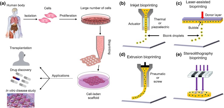
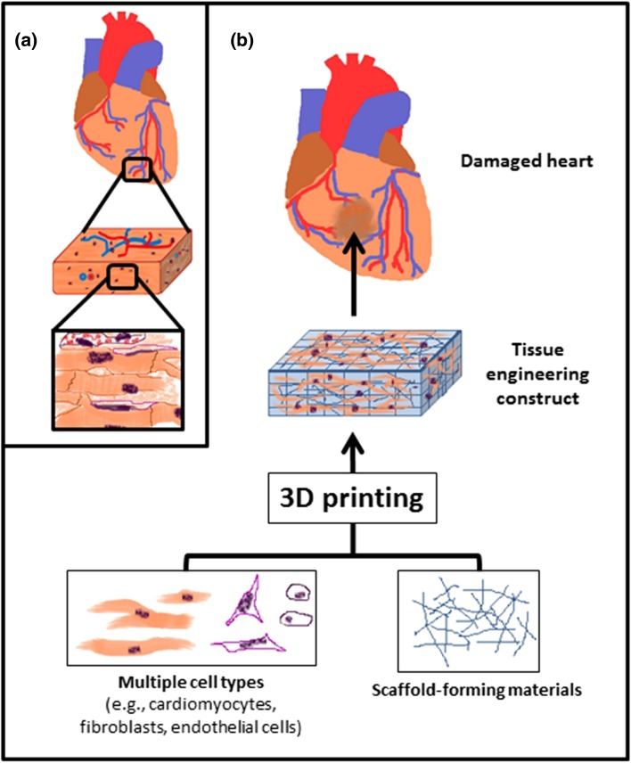
Stem cells offer tremendous promise for regenerative medicine as they can become a variety of cell types. They also continuously proliferate, providing a renewable source of cells. Recently, it has been found that 3D printing constructs using stem cells, can generate models representing healthy or diseased tissues, as well as substitutes for diseased and damaged tissues. Here, we review the current state of the field of 3D printing stem cell derived tissues. First, we cover 3D printing technologies and discuss the different types of stem cells used for tissue engineering applications. We then detail the properties required for the bioinks used when printing viable tissues from stem cells. We give relevant examples of such bioprinted tissues, including adipose tissue, blood vessels, bone, cardiac tissue, cartilage, heart valves, liver, muscle, neural tissue, and pancreas. Finally, we provide future directions for improving the current technologies, along with areas of focus for future work to translate these exciting technologies into clinical applications.

Keywords: Bioinks; Biomaterials; Controlled; Drug delivery; Pluripotent stem cells; Regenerative medicine; Stem cell niche; Tissue engineering.


Figures

Figure 1
Figure 1
Bioprinting process, techniques, and applications. (a) For human therapeutic applications, the typical workflow of bioprinting would involve the isolation and expansion of human cells prior to printing the desired cell-laden scaffold. These scaffolds could then ultimately be used as therapeutic devices themselves, as a testing platform for drug screening and discovery, or as an in vitro model system for disease. (b) Inkjet printers eject small droplets of cells and hydrogel sequentially to build up tissues. (c) Laser bioprinters use a laser to vaporize a region in the donor layer (top) forming a bubble that propels a suspended bioink to fall onto the substrate. (d) Extrusion bioprinters use pneumatics or manual force to continuously extrude a liquid cell–hydrogel solution. (e) Stereolithographic printers use a digital light projector to selectively crosslink bioinks plane-by-plane. In (c) and (e), colored arrows represent a laser pulse or projected light, respectively (adapted from with permission from Ref. 121).
Figure 2
Figure 2
Hollow channels can be created in multiple geometries using a fugitive bioink and then subsequently perfusing the system with a liquid or cell-laden suspension (adapted from with permission from Ref. 99).
Figure 3
Figure 3
Microextrusion-printed grid structures based on cell-laden HAp-bioink. 3D grid structure with a height of 5 mm and edge length of 10 mm; (a) printing with an extrusion-based printing system and (b) after 24 h incubation in DMEM under physiologic conditions. Light microscopic evaluation of a cross-section of the grid structure; (c) directly after printing and (d) after incubation. (e) Fluorescence microscope evaluation of cell distribution in the printed structure after 24 h of culture (adapted from Ref. 178).
Figure 4
Figure 4
Schematic representation of 3D printing for tissue engineering applications, such as for cardiac tissue engineering. (a) Tissues are composed of multiple types of cells assembled into hierarchal structures. (b) 3D printing can be utilized to assemble functional tissue from cells and scaffold-forming materials (adopted with permission from Ref. 150).
Figure 5
Figure 5
Handheld biopen developed for in situ cartilage tissue printing. (a) Schematic illustration of handheld biopen. (b)Schematic representation of coaxial nozzle. (c) Picture of the cartridges dedicated to Core and Shell loading in the printer, with relative magnification of the nozzle during co-axial deposition. (d) Representative 3D rendered confocal images of Core/Shell printed sample labeled with fluorescent beads (adapted from Ref. licensed under http://creativecommons.org/licenses/by/4.0/).
Figure 6
Figure 6
Fabrication of biomimetic nanofibrous scaffolds for neural tissue engineering. (a) Strategies of electrospinning and self-assembly used for nanofiber fabrication. (b) A general pathway of engineering neural tissues with the use of nerve cells and nanofibers composed of natural polymers and synthetic polymers. (c) Schematic illustration and image of neural cells on nanofibrous scaffolds (adapted with permission from Ref. licensed under http://creativecommons.org/licenses/by/4.0/).

References

    1. AlGhamdi KM, Kumar A, Moussa NA. Low-level laser therapy: a useful technique for enhancing the proliferation of various cultured cells. Lasers Med. Sci. 2012;27(1):237–249. - PubMed
    1. Ali M, et al. Controlling laser-induced jet formation for bioprinting mesenchymal stem cells with high viability and high resolution. Biofabrication. 2014;6(4):045001. - PubMed
    1. Anil Kumar, S.A.K., S. Park, Y. Ito, B. Joddar. Photo-crosslinkable furfurl-gelatin as a novel bioink for 3D bioprinting of cardiac tissue. In: Annual Meeting of the BMES, 2017, Phoenix, Arizona, 2017.
    1. AsteriasBiotherapeutics, Asterias Biotherapeutics Announces Dosing of First Patient in New SCiSTAR Clinical Trial Cohort Testing AST-OPC1 in an Expanded Cervical Spinal Cord Injury Patient Population. 2016. Asteriasbiotherapuetics.com.
    1. Bajaj P, et al. Patterned three-dimensional encapsulation of embryonic stem cells using dielectrophoresis and stereolithography. Adv. Healthc. Mater. 2013;2(3):450–458. - PubMed

LinkOut - more resources