

Tracheal granular cell tumour presenting with throat discomfort
- PMID: 31719987
- PMCID: PMC6837852
- DOI: 10.1002/rcr2.499
Tracheal granular cell tumour presenting with throat discomfort
Abstract
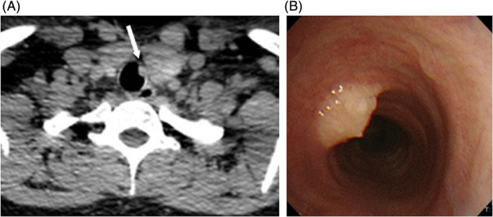
Granular cell tumours (GCTs) are a rare form of neoplasm found throughout the body. Tracheobronchial involvement is less common. We describe a case of tracheal GCT in a 37-year-old Japanese woman presenting with throat discomfort. A tracheal tumour was found during laryngoscopy for undefined throat discomfort. Bronchoscopy demonstrated a white sub-epithelial solitary nodule on the tracheal wall, and pathological examination of the biopsy samples confirmed GCT. No therapeutic procedures were performed, and the tumour is currently under strict observation. Throat discomfort is a rare presentation of tracheal tumours, but an early inspection using laryngoscopy and bronchoscopy may be helpful in determining an accurate diagnosis.
Keywords: Bronchoscopy; granular cell tumour; throat discomfort; tracheal tumour.
© 2019 The Authors. Respirology Case Reports published by John Wiley & Sons Australia, Ltd on behalf of The Asian Pacific Society of Respirology.
Figures

Similar articles
-
Subcutaneous granular cell tumour of the lumbar region.J Cutan Aesthet Surg. 2011 May;4(2):132-4. doi: 10.4103/0974-2077.85039. J Cutan Aesthet Surg. 2011. PMID: 21976906 Free PMC article.
-
[Granular cell tumour in a patient with pulmonary tuberculosis].Pneumologie. 2008 Mar;62(3):158-61. doi: 10.1055/s-2007-996163. Epub 2008 Jan 16. Pneumologie. 2008. PMID: 18200457 German.
-
Granular cell tumor of the trachea mimicking an infiltrating thyroid cancer. A case report.Int J Surg Case Rep. 2022 May;94:107031. doi: 10.1016/j.ijscr.2022.107031. Epub 2022 Apr 6. Int J Surg Case Rep. 2022. PMID: 35398784 Free PMC article.
-
Management of pediatric airway granular cell tumor: role of laryngotracheal reconstruction.Int J Pediatr Otorhinolaryngol. 2006 Jun;70(6):957-63. doi: 10.1016/j.ijporl.2005.12.018. Epub 2006 Feb 8. Int J Pediatr Otorhinolaryngol. 2006. PMID: 16466812 Review.
-
Solitary fibrous tumor of the trachea: a case report.Gen Thorac Cardiovasc Surg. 2020 Dec;68(12):1523-1527. doi: 10.1007/s11748-019-01274-5. Epub 2019 Dec 17. Gen Thorac Cardiovasc Surg. 2020. PMID: 31848901 Free PMC article. Review.
References
-
- Van der Maten J, Blaauwgeers JL, Sutedja TG, et al. 2003. Granular cell tumors of the tracheobronchial tree. J. Thorac. Cardiovasc. Surg. 126:740–743. - PubMed
-
- Ipakchi R, Zager WH, de Baca ME, et al. 2004. Granular cell tumor of the trachea in pregnancy: a case report and review of literature. Laryngoscope 114:143–147. - PubMed
Publication types
LinkOut - more resources
Full Text Sources
