. 2019 Oct 17;19(5):e34.
doi: 10.4110/in.2019.19.e34. eCollection 2019 Oct.

Neutrophilic Granule Protein Is a Novel Murine LPS Antagonist

Affiliations

Neutrophilic Granule Protein Is a Novel Murine LPS Antagonist

Jaewoo Hong et al. Immune Netw. .

Abstract

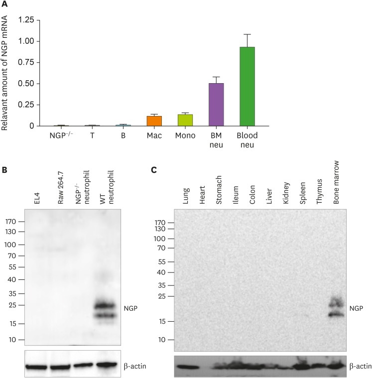
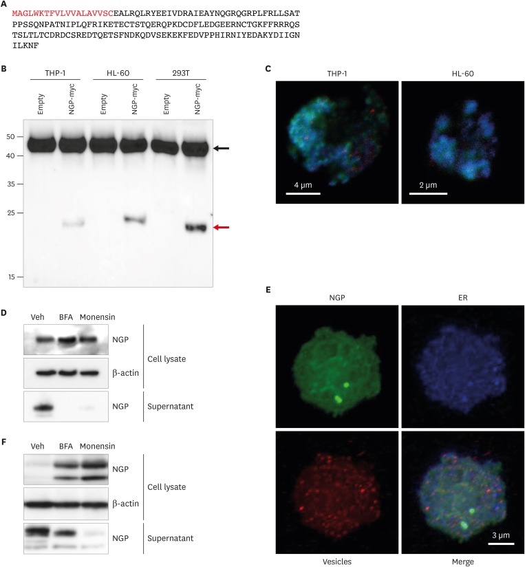
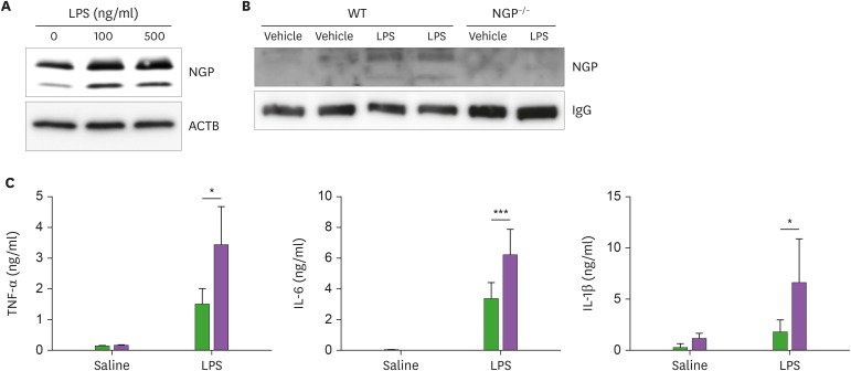
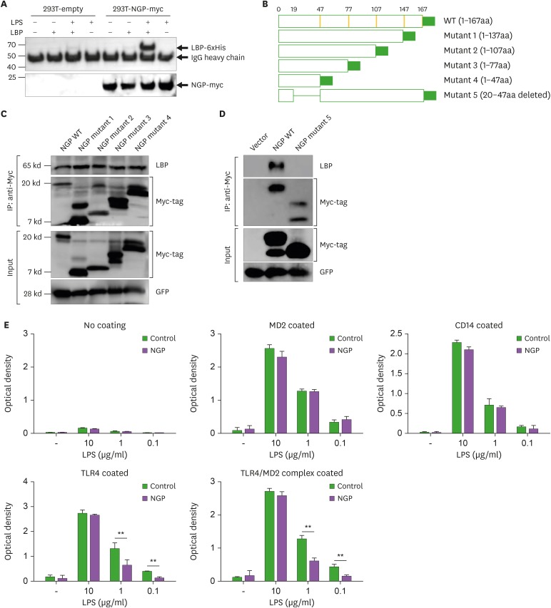
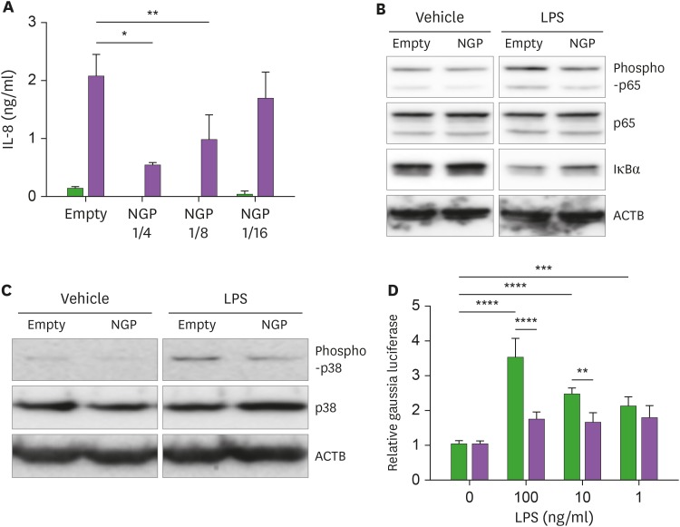
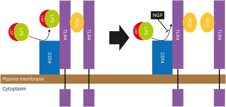
Neutrophilic granule protein (NGP) was previously reported as a granular protein of neutrophils in mouse, but the function has not been known clearly. We found the presence of the possible signal peptide in NGP and validated this protein is circulating in the bloodstream. In our findings, NGP is being modified post-translationally in Golgi apparatus and endoplasmic reticulum, which is a universal character of secretory molecules with a signal peptide. The secreted NGP protein could be detected both in vitro and in vivo. NGP has sequence similarity with an antimicrobial protein cathelicidin, and we observed the aspect of inflammation of NGP. Interestingly, NGP interacts with the complex of LPS and LPS binding protein (LBP). This interaction blocks the binding of the complex of LPS and LBP to TLR4 and the downstream inflammatory signals. Furthermore, the inhibitory function of NGP against the inflammatory effect of LPS could be observed in both in vitro and in vivo. With these findings, we report NGP is a novel secretory protein to mask LPS and inhibit its function.

Keywords: Cytokines; Inflammation; Inhibitors; Lipopolysaccharides; Neutrophil; Protein binding.


Conflict of interest statement

Conflicts of Interest: The authors declare no potential conflicts of interest.

Figures

Figure 1
Figure 1. NGP is expressed specifically in mouse neutrophils. (A) T cells, B cells, macrophages, monocytes, BM-derived neutrophils, and circulating neutrophils were isolated from a WT mouse. mRNA level of NGP was relatively measured by qRT-PCR and compared to NGP deficient neutrophils as the negative control and blood neutrophil as the positive control (n=3, quadruplet mean±SD). (B) NGP protein was detected by Western blotting from the total lysate of EL4 cells, Raw 264.7 cells, NGP deficient neutrophils, and WT neutrophils by Western blot. (C) NGP protein was detected by Western blotting from the total lysate of mouse tissues. Lung, heart, stomach, ileum, colon, liver, kidney, spleen, thymus, and BM were collected from a WT mouse. Tissue was ground in 1× sample buffer, and NGP was detected by Western blotting.
Figure 2
Figure 2. NGP is a soluble secretory molecule. (A) The amino acid sequence of NGP. Red letters stand for the area of the signal peptide (red). (B) Myc-tagged NGP was pulled down from NGP overexpressed THP-1, HL-60, or 293T cells. Myc-tag was detected by Western blotting, and the red arrow shows NGP-Myc band. The black arrow shows IgG heavy chain. Representative images are shown. (C) NGP overexpressing HL-60 (upper panel), and THP-1 (lower panel) were stained against Golga3 (red), Calreticulin (green), and NGP (blue). The stained cells were analyzed by confocal microscopy. (D) NGP-Myc overexpressing THP-1 cells were treated by veh, 1× BFA, or 1× monensin (Biolegend) for 3 h. Myc-tagged NGP was detected from the total lysate and supernatant by Western blotting. A representative result out of 3 experiments is shown. (E) Mouse peritoneal neutrophils were stained against NGP (green), Calreticulin (blue), and lysobrite for granules (red). The stained cells were analyzed by confocal microscopy. A representative image is shown. (F) Mouse peritoneal neutrophils were treated by veh, 1× BFA, or 1× monensin (Biolegend) for 3 h. NGP was detected from the total lysate and supernatant by Western blotting. A representative result out of 3 experiments is shown.
Veh, vehicle.
Figure 3
Figure 3. NGP is induced by LPS. (A) Mouse peritoneal neutrophils were treated by vehicle, 100 ng/ml, or 500 ng/ml of LPS for 3 h. NGP was detected from the total lysate by Western blotting. (B) 1 mg/kg of LPS was injected into WT mouse or NGP deficient mouse intraperitoneally. The serum was isolated from each animal 5 h after injection. NGP was probed from the serum by Western blotting. (C) 0.2 mg/ml of LPS was injected into WT (black bar) or NGP deficient (empty bar) mice. Mouse serum was collected before injection of LPS, 1 h after injection for detection of IL-1β and IL-6, and 3 h after injection to detect TNFα. TNFα, IL-1β, and IL-6 were detected by ELISA (quadruplet mean±SD). Each experiment was repeated 3 times, and representative data is selected.
*p<0.05, ***p<0.001.
Figure 4
Figure 4. NGP masks LPS and inhibits LPS receptor binding. (A) Conditional medium of NGP or empty vector was mixed with 100 ng of recombinant LBP-His and/or 100 ng of LPS for 30 min on ice. LBP or NGP was detected by Western blotting. (B) Five different NGP mutant constructs were prepared to identify the binding site of LPS-LBP complex on NGP. Amino acids are deleted from the C-terminus in mutant 1–4 as designated. Mutant 5 has a deletion of 28 amino acids after signal peptide. (C and D) WT NGP, or deletion mutants of NGP were mixed with 100 ng of LPS and 100 ng of LBP for 30 min on ice. NGP was pulled down with Myc-tagged NGP, and LBP and Myc-tagged NGP were detected by Western blotting from both IP products and input. (E) Recombinant MD2, CD14, TLR4, or TLR4/MD2 complex was coated on an ELISA plate. Biotinylated LPS was mixed with 100 ng of LBP in designated concentration for 30 min on ice. The mixture of LPS and LBP was added to the receptor coated plate for 2 h. Bound LPS was visualized by TMB solution and analyzed by ELISA reader (quadruplet mean±SD).
GFP, green fluorescent protein. **p<0.01.
Figure 5
Figure 5. NGP blocks the activity of LPS. (A) THP-1 cells were treated by vehicle (black bars) or 100 ng/ml of LPS (blank bars) for 16 h in the mixture of NGP conditional medium and empty vector conditional medium in a ratio of 0, 1/4, 1/8, and 1/16. IL-8 was detected from the culture medium (quadruplet mean±SD). (B) THP-1 cells were treated by vehicle or 10 ng/ml of LPS in an empty vector or NGP conditional medium for 30 min. Phospho-NF-κB p65, NF-κB p65, and IκBα were detected by Western blotting. (C) THP-1 cells were treated by vehicle or 10 ng/ml of LPS in an empty vector or NGP conditional medium for 30 min. Phospho-p38 MAPK and p38 MAPK were detected by Western blotting. (D) The conditional medium of empty vector (black) or NGP (blank) was treated overnight. LPS was mixed in different concentrations and then treated to 293 cells transfected with Gaussia luciferase NF-κB construct. The luminescence of cells with NGP medium showed inhibited activity of the reporter (quadruplet mean±SD).
*p<0.05, **p<0.01, ***p<0.001, ****p<0.0001.
Figure 6
Figure 6. NGP masks LPS-LBP complex. The complex of LPS and LBP stimulates the LPS receptor complex of CD14, TLR4, and MD2. NGP binds to the LPS-LBP complex and masks the complex to interact with its receptor. LPS-LBP complex fails to bind to TLR4 from CD14 and followed by the null downstream signal of TLR4.

References

    1. Gazzano-Santoro H, Parent JB, Grinna L, Horwitz A, Parsons T, Theofan G, Elsbach P, Weiss J, Conlon PJ. High-affinity binding of the bactericidal/permeability-increasing protein and a recombinant amino-terminal fragment to the lipid A region of lipopolysaccharide. Infect Immun. 1992;60:4754–4761. - PMC - PubMed
    1. Shafer WM, Martin LE, Spitznagel JK. Cationic antimicrobial proteins isolated from human neutrophil granulocytes in the presence of diisopropyl fluorophosphate. Infect Immun. 1984;45:29–35. - PMC - PubMed
    1. Lehrer RI, Lichtenstein AK, Ganz T. Defensins: antimicrobial and cytotoxic peptides of mammalian cells. Annu Rev Immunol. 1993;11:105–128. - PubMed
    1. Pereira HA, Erdem I, Pohl J, Spitznagel JK. Synthetic bactericidal peptide based on CAP37: a 37-kDa human neutrophil granule-associated cationic antimicrobial protein chemotactic for monocytes. Proc Natl Acad Sci U S A. 1993;90:4733–4737. - PMC - PubMed
    1. Hirata M, Yoshida M, Inada K, Kirikae T. Investigation of endotoxin binding cationic proteins from granulocytes; agglutination of erythrocytes sensitized with Re-LPS. Adv Exp Med Biol. 1990;256:287–299. - PubMed