. 2019 Oct 24;4(19):18342-18354.
doi: 10.1021/acsomega.9b02588. eCollection 2019 Nov 5.

Hydrophilic Fluorescent Nanoprodrug of Paclitaxel for Glioblastoma Chemotherapy

Affiliations

Hydrophilic Fluorescent Nanoprodrug of Paclitaxel for Glioblastoma Chemotherapy

Jonathan Daniel et al. ACS Omega. .

Abstract

Highly water-soluble, nontoxic organic nanoparticles on which paclitaxel (PTX), a hydrophobic anticancer drug, has been covalently bound via an ester linkage (4.5% of total weight) have been prepared for the treatment of glioblastoma. These soft fluorescent organic nanoparticles (FONPs), obtained from citric acid and diethylenetriamine by microwave-assisted condensation, show suitable size (Ø = 17-30 nm), remarkable solubility in water, softness as well as strong blue fluorescence in an aqueous environment that are fully retained in cell culture medium. Moreover, these FONPs were demonstrated to show in vitro safety and preferential internalization in glioblastoma cells through caveolin/lipid raft-mediated endocytosis. The PTX-conjugated FONPs retain excellent solubility in water and remain stable in water (no leaching), while they showed anticancer activity against glioblastoma cells in two-dimensional and three-dimensional culture. PTX-specific effects on microtubules reveal that PTX is intracellularly released from the nanocarriers in its active form, in relation with an intracellular-promoted lysis of the ester linkage. As such, these hydrophilic prodrug formulations hold major promise as biocompatible nanotools for drug delivery.


Conflict of interest statement

The authors declare no competing financial interest.

Figures

Scheme 1
Scheme 1. Synthesis Steps of the FONP–PTX Prodrug
Step 1: Synthesis of FONP platforms. Citric acid, diethylenetriamine, water, microwave (600 W), 2 min; step 2: Activation of FONP platforms. Succinic anhydride, Na2CO3, DMSO, rt, 2 h; step 3: Grafting of PTX. EDC·HCl, DMAP, PTX, DMF, rt, 60 h.
Figure 1
Figure 1
(A) TEM images of FONPs with size distribution fitted with a log normal in caption; (B) TEM images of FONPs–PTX with size distribution fitted with a log normal in caption; (C) AFM images of FONPs; (D) 1H NMR spectrum in DMSO-d6 of FONPs–PTX; (E) IR spectrum of FONPs; and (F) carbon XPS spectrum of FONPs.
Figure 2
Figure 2
(A) Normalized absorption (purple) and emission (cyan) spectra of nanoparticles in water; (B) normalized three-dimensional (3D) excitation and emission spectra of nanoparticles in water; and (C) 2PA spectrum (black) compared to rescaled one-photon absorption spectrum (red) of FONPs in water.
Figure 3
Figure 3
Colorimetric cell survival assays performed after 72 h of incubation of FONPs (1–100 μg/mL) with U-87 MG cells (A), HMEC-1 (B), and NHDF (C) and the real-time impedance-based survival assay performed over a period of 72 h after treatment with FONPs (5–100 μg/mL) for U-87 MG cells (D), HMEC-1 (E), and NHDF (F). MTT: 3-(4,5-dimethylthiazol-2-yl)-2,5-diphenyltetrazolium bromide.
Figure 4
Figure 4
Two-photon fluorescence images of U-87 MG cells (upper panel) and HMEC-1 and NHDF (lower panel) after incubation for 4 h with FONPs (1 or 5 μg/mL), upon excitation at 740 nm and emission detection in the 480–550 nm range, after autofluorescence correction. Orthogonal views showing the distribution of FONPs in the thickness of the cells are presented for U-87 MG cells (middle panel). Transmission images are provided to locate the cells. Scale bar: 35 μm.
Figure 5
Figure 5
Inhibition of cell viability by FONPs–PTX in 2D glioblastoma cell culture. (A) MTT assay on U-87 MG cells treated with FONPs–PTX for 72 h. (B) Immunofluorescence imaging of the microtubular network and nuclei (40×) in U-87 MG cells untreated (control) or incubated with FONPs–PTX (5 or 10 μM) for 24 h. Bundles (full arrow), pseudo-asters (dotted arrow), and mitotic block are typical of the PTX pharmacological effect.
Figure 6
Figure 6
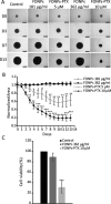
Safety of FONPs and anticancer activity of FONPs–PTX in 3D cell culture. (A) Phase-contrast microscopy (4×) of U-87 MG spheroid control and treated with FONPs or FONPs–PTX for 13 days (scale bar = 500 μm, D = day). (B) Time- and dose-dependent effect of FONPs–PTX and absence of activity of FONPs on the area of spheroids (normalized area to untreated control spheroids) until 13 days. (C) Alamar blue assay on spheroids after 13 days of treatment with FONPs (362 μg/mL) or FONPs–PTX (10 μM) compared to untreated control spheroids.

References

    1. Jain V.; Jain S.; Mahajan S. C. Nanomedicines Based Drug Delivery Systems for Anti-Cancer Targeting and Treatment. Curr. Drug Deliv. 2015, 12, 177–191. 10.2174/1567201811666140822112516. - DOI - PubMed
    1. Sun T.; Zhang Y. S.; Pang B.; Hyun D. C.; Yang M.; Xia Y. Engineered Nanoparticles for Drug Delivery in Cancer Therapy. Angew. Chem. Int. Ed. Engl. 2014, 53, 12320–12364. 10.1002/anie.201403036. - DOI - PubMed
    1. Pérez-Herrero E.; Fernández-Medarde A. Advanced Targeted Therapies in Cancer: Drug Nanocarriers, the Future of Chemotherapy. Eur. J. Pharm. Biopharm. 2015, 93, 52–79. 10.1016/j.ejpb.2015.03.018. - DOI - PubMed
    1. Nabil G.; Bhise K.; Sau S.; Atef M.; El-Banna H. A.; Iyer A. K. Nano-Engineered Delivery Systems for Cancer Imaging and Therapy: Recent Advances, Future Direction and Patent Evaluation. Drug Discov. Today 2019, 24, 462–491. 10.1016/j.drudis.2018.08.009. - DOI - PMC - PubMed
    1. Kim S.-W.; Lee Y. K.; Kim S.-H.; Park J.-Y.; Lee D. U.; Choi J.; Hong J. H.; Kim S.; Khang D. Covalent, Non-Covalent, Encapsulated Nanodrug Regulate the Fate of Intra- and Extracellular Trafficking: Impact on Cancer and Normal Cells. Sci. Rep. 2017, 7, 6454.10.1038/s41598-017-06796-7. - DOI - PMC - PubMed

LinkOut - more resources