

Contrast-enhanced echocardiographic measurement of longitudinal strain: accuracy and its relationship with image quality
- PMID: 31720940
- PMCID: PMC7316528
- DOI: 10.1007/s10554-019-01732-4
Contrast-enhanced echocardiographic measurement of longitudinal strain: accuracy and its relationship with image quality
Abstract
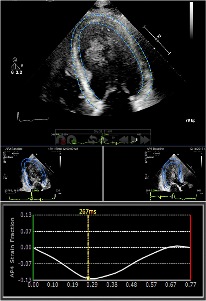
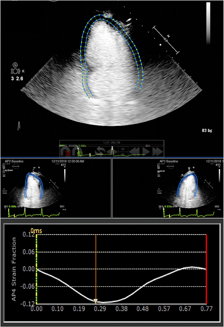
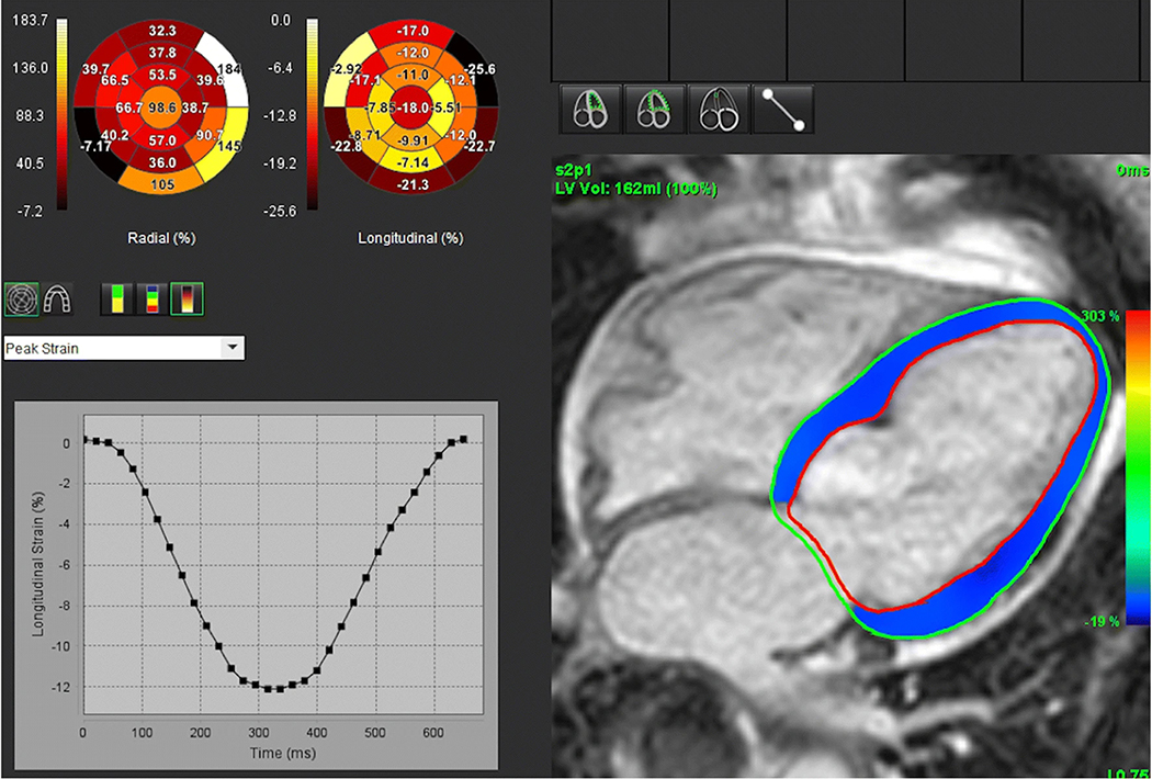
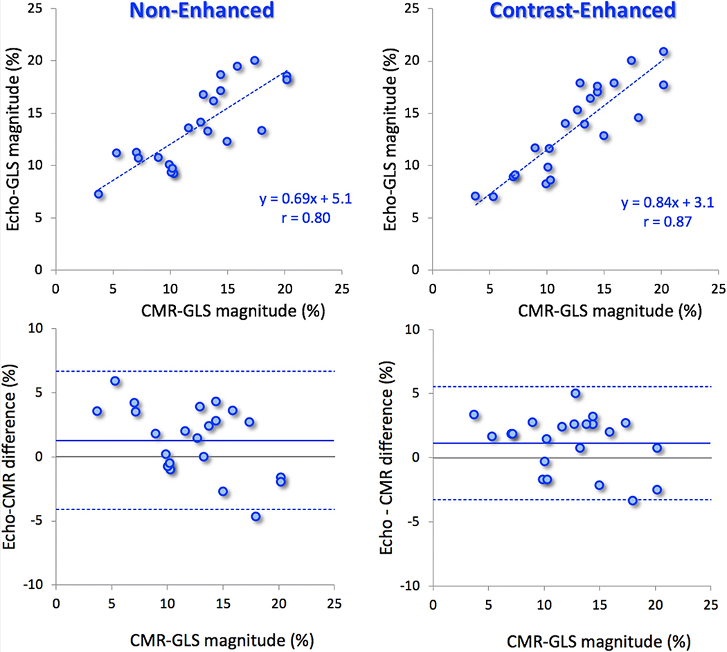
The importance of left ventricular (LV) global longitudinal strain (GLS) is increasingly recognized in multiple clinical scenarios. However, in patients with poor image quality, strain is difficult or impossible to measure without contrast enhancement. The feasibility of contrast-enhanced GLS measurement was recently demonstrated. We sought to determine: (1) whether contrast enhancement improves the accuracy of GLS measurements against cardiac magnetic resonance (CMR) reference, (2) their reproducibility compared to non-enhanced GLS, and (3) the dependence of accuracy and reproducibility on image quality. We prospectively enrolled 25 patients undergoing clinically indicated CMR imaging who subsequently underwent transthoracic echocardiography (TTE) with and without low-dose contrast injection (1-2 mL Optison/3-5 mL saline IV, GE Healthcare). GLS was measured from both non-contrast and contrast-enhanced images using speckle tracking (EchoInsight, Epsilon Imaging). These measurements were compared to each other and to CMR reference values obtained using feature tracking (SuiteHEART, NeoSoft). Inter-technique comparisons included linear regression and Bland-Altman analyses. A random subgroup of 15 patients was used to assess inter- and intra-observer variability using intra-class correlation (ICC). Contrast-enhanced GLS was in close agreement with non-enhanced GLS (r = 0.95; bias: - 0.2 ± 1.5%). Both inter-observer (ICC = 0.88 vs. 0.82) and intra-observer variability (ICC = 0.91 vs. 0.88) were improved by contrast enhancement. The agreement with CMR was better for contrast-enhanced GLS (r = 0.87; bias: 1.1 ± 2.2%) than for non-enhanced GLS (r = 0.80; bias: 1.3 ± 2.7%). In 12/25 patients with suboptimal TTE images that rendered GLS difficult to measure, contrast-enhanced GLS showed better agreement with CMR than non-enhanced GLS (r = 0.88 vs. 0.83) and also improved inter-observer (ICC = 0.83 vs. 0.76) and intra-observer variability (ICC = 0.88 vs. 0.82). In conclusion, contrast enhancement of TTE images improves the accuracy and reproducibility of GLS measurements, resulting in better agreement with CMR, even in patients with suboptimal acoustic windows. This approach may aid in the assessment of LV function in this patient population.
Keywords: Contrast enhancement; Left ventricular function; Myocardial strain; Speckle-tracking echocardiography.
Conflict of interest statement
Compliance with ethical standards
Figures
References
-
- Cho GY, Marwick TH, Kim HS, Kim MK, Hong KS, Oh DJ (2009) Global 2-dimensional strain as a new prognosticator in patients with heart failure. J Am Coll Cardiol 54:618–624 - PubMed
-
- Leitman M, Lysyansky P, Sidenko S, Shir V, Peleg E, Binenbaum M et al. (2004) Two-dimensional strain-a novel software for real-time quantitative echocardiographic assessment of myocardial function. J Am Soc Echocardiogr 17:1021–1029 - PubMed
-
- Mignot A, Donal E, Zaroui A, Reant P, Salem A, Hamon C et al. (2010) Global longitudinal strain as a major predictor of cardiac events in patients with depressed left ventricular function: a multicenter study. J Am Soc Echocardiogr 23:1019–1024 - PubMed
-
- Serri K, Reant P, Lafitte M, Berhouet M, Le Bouffos V, Roudaut R et al. (2006) Global and regional myocardial function quantification by two-dimensional strain: application in hypertrophic cardiomyopathy. J Am Coll Cardiol 47:1175–1181 - PubMed
-
- Stanton T, Leano R, Marwick TH (2009) Prediction of all-cause mortality from global longitudinal speckle strain: comparison with ejection fraction and wall motion scoring. Circ Cardiovasc Imaging 2:356–364 - PubMed
Publication types
MeSH terms
Substances
Grants and funding
LinkOut - more resources
Full Text Sources
Medical
Miscellaneous
