. 2020 Feb;38(2):301-314.
doi: 10.1002/stem.3117. Epub 2019 Nov 26.

Hes1 regulates anagen initiation and hair follicle regeneration through modulation of hedgehog signaling

Affiliations

Hes1 regulates anagen initiation and hair follicle regeneration through modulation of hedgehog signaling

Wei-Jeng Suen et al. Stem Cells. 2020 Feb.

Abstract

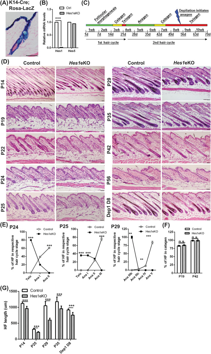
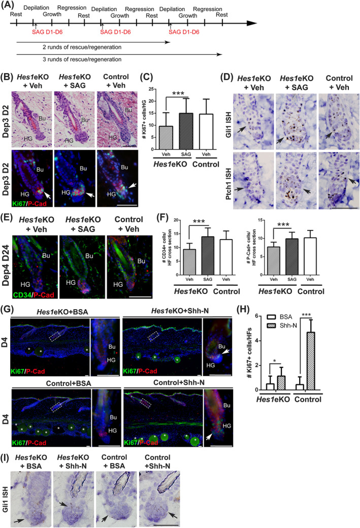
Adult hair follicles undergo repeated cycling of regression (catagen), resting (telogen), and growth (anagen), which is maintained by hair follicle stem cells (HFSCs). The mechanism underlying hair growth initiation and HFSC maintenance is not fully understood. Here, by epithelial deletion of Hes1, a major Notch downstream transcriptional repressor, we found that hair growth is retarded, but the hair cycle progresses normally. Hes1 is specifically upregulated in the lower bulge/HG during anagen initiation. Accordingly, loss of Hes1 results in delayed activation of the secondary hair germ (HG) and shortened anagen phase. This developmental delay causes reduced hair shaft length but not identity changes in follicular lineages. Remarkably, Hes1 ablation results in impaired hair regeneration upon repetitive depilation. Microarray gene profiling on HFSCs indicates that Hes1 modulates Shh responsiveness in anagen initiation. Using primary keratinocyte cultures, we demonstrated that Hes1 deletion negatively influences ciliogenesis and Smoothened ciliary accumulation upon Shh treatment. Furthermore, transient application of Smoothened agonist during repetitive depilation can rescue anagen initiation and HFSC self-renewal in Hes1-deficient hair follicles. We reveal a critical function of Hes1 in potentiating Shh signaling in anagen initiation, which allows sufficient signaling strength to expand the HG and replenish HFSCs to maintain the hair cycle homeostasis.

Keywords: adult stem cells; cellular proliferation; epidermis; notch; signal transduction.


Conflict of interest statement

The authors indicated no potential conflicts of interest.

Figures

Figure 1
Figure 1
Hes1 deletion using K14‐Cre deleter line causes retarded hair growth. A, X‐gal‐stained back skin section of [K14‐Cre+/wt; Rosa‐LacZ +/wt] mice with eosin counterstain. B, Quantitative real‐time PCR (qRT‐PCR) analysis of Hes1 and Hes5 expression levels in the back skin epithelium of Hes1eKO and control mice (mean ± SD, n = 4, ***P < .001). C, Illustration of the two synchronous hair cycles coordinated with age in mice after birth. D, Hematoxylin and eosin‐stained back skin sections at P14 (follicular morphogenesis), P19 (first catagen), P22 (first telogen), P24 (second early anagen), P25 (second anagen), P29 (second anagen), P35 (second late‐anagen/early catagen), P42 (second catagen), P56 (second telogen), and day 8 post‐depilation at P56. E and F, Quantification of hair cycle stage according to morphology‐based hair cycle histomorphometry25 in each hair cycle phase as indicated (mean ± SD, n > 50 hair follicles (HFs) per genotype from two to three independent control and mutant pairs at each phase, n.s., nonsignificant; **P < .01; ***P < .001). G, Quantification of HF length (from the base of follicle to the follicular orifice) in each hair cycle phase as indicated (mean ± SD, n > 50 HFs per genotype from two to three independent control and mutant pairs at each phase, ***P < .001). Scale bar, 50 μm
Figure 2
Figure 2
Hes1 deletion results in delayed anagen initiation and shortened hair follicle (HF) growth phase. A, In situ hybridization of Hes1 (arrows) in back skin sections at P22 and P24 with hematoxylin counterstain. The dotted lines demarcate the bulge and the solid lines demarcate the boundary between DP and HG when visible. B, Back skin sections were immunostained for Hes1 at P22 and P24. C, Back skin sections were immunostained for HG marker P‐Cadherin (P‐Cad, arrows) at P22. D, Quantification of P‐Cad + cells in the HF at P22 (mean ± SD, n > 30 HFs per genotype from three independent control and mutant pairs, n.s., nonsignificant). E, Back skin sections were double immunostained for P‐Cad and proliferative marker Ki67 (arrows) at P24. F, Quantification of Ki67+ cells in the P‐Cad + cells at P24 (mean ± SD, n > 30 HFs per genotype from three independent control and mutant pairs, ***P < .001). G, Back skin sections at P24 were immunostained for β‐catenin. The arrows mark the nuclear β‐catenin staining. The dotted lines denote the boundary between DP and HG when visible. H, Back skin sections immunostained for phospho‐Smad1/5/8 (arrows). The dotted line marks the HF and solid line marks the DP. I, Double immunostaining of AE13 and KI67 in back skin sections at P29 (full anagen) and P35 (late anagen/early catagen). The dotted lines depict the line of Auber. J, Quantification of the bulb size (Ki67+ area below the line of Auber) (mean ± SD, >40 HFs from three biological replicates per genotype per stage, ***P < .001). K, Back skin sections immunostained for cell mitotic marker phospho‐histone H3 (p‐H3) at P29 and P35. L, Quantification of p‐H3+ cells in the hair matrix (mean ± SD, >40 HFs from three biological replicates per genotype per stage, ***P < .001). DAPI counterstaining in blue. Bu, bulge; HG, hair germ; DP, derma papillae; Bb, hair bulb. M, Bright field images of club hair of four different hair types at P60. N, Quantification of club hair length of each HF type (mean ± SD, 20 club hairs for each hair types per mouse, n = 3 biological replicates per genotype, ***P < .001 determined by analysis of variance). Scale bar, 50 μm except (M), where it is 1 mm
Figure 3
Figure 3
Hes1 deficiency causes compromised hair follicle (HF) regeneration and hair follicle stem cell (HFSC) self‐renewal after repetitive depilation. A, Sequential depilation of control littermate (Ctrl) and Hes1 conditional knockout (Hes1eKO) mice for four rounds with a three‐week interval from the second telogen. Representative pictures of male mice are shown (n = 5). B, Close up of back skin at day 22 post‐depilation‐induced hair regeneration. C, Back skin sections from repetitive depilation (day 22 after fourth depilation) were double immunostained for CD34 and P‐Cad. D, Quantification of CD34+ bulge and P‐Cad + HG cells in HFs after four rounds of sequential depilation (mean ± SD, n > 50 HFs per genotype from four independent control and mutant pairs, ***P < .001). E, Quantifications of HFSCs after four rounds of sequential depilation using flow cytometry. Data are presented as percentage of Bu/CD49f + cells relative to control samples (mean ± SD, n = 4 independent pairs). F, Skin sections from sequential depilation (one and five times) were processed for CD34 immunostaining and EdU incorporation assays. Dotted lines denote the DP when visible. G, Quantification of cell proliferation within CD34+ cells and below CD34+ cells after sequential depilation (mean ± SD, n > 50 HFs per genotype from three independent control and mutant pairs for each set, ***P < .001). Bu, Bulge; HG, hair germ; SG, sebaceous glands. DAPI counterstaining in blue. Scale bar, 50 μm
Figure 4
Figure 4
Hes1 inactivation causes compromised Shh signaling in hair follicle stem cells (HFSCs). A, Ingenuity pathway analysis result showing the top diseases and bio functions and top canonical pathways significantly affected by Hes1 deletion in HFSCs (cutoff fold change >1.5 or <−1.5, P < .05). Red and green indicate upregulated and downregulated genes in Hes1eKO HFSCs, respectively. B, Gene set enrichment analysis of microarray gene expression profiling on HFSCs from control and Hes1eKO mice (n = 2 independent pairs). C and D, qRT‐PCR analysis of selected genes related to Shh signaling, top diseases and biological functions, and top canonical pathways on FACS‐purified HFSCs from control and Hes1eKO mice at P72 (telogen after depilation at P50, mean ± SD, n = 4 independent pairs, *P < .05, **P < .01, ***P < .001). E, In situ hybridization of Gli1 and Ptch1 (arrows) in control and Hes1eKO back skin sections at P72 (telogen after depilation at P50). Scale bar, 50 μm
Figure 5
Figure 5
Hes1 deletion causes primary cilia defect and influences Shh signaling responsiveness. A, Double immunostaining of Arl13b and Pericentrin in back skin sections of mice at P72 (telogen after depilation at P50). The dotted line box denotes the region of cilia measurement. The boxed region shows high magnification. DAPI counterstaining in blue. B, Quantification of cilia length in lower hair follicles (HFs) (dotted line box in (A)) of control and Hes1eKO mice (mean ± SD, n = 3 independent pairs; control 336 cilia; Hes1eKO 384 cilia, ***P < .01). C, Control and Hes1eKO PMEKs were immunostained for Arl13b and Pericentrin. The boxed region shows high magnification. D, Quantification of ciliated cells expressed as a percentage of total in control and Hes1eKO PMEKs (mean ± SD, n = 3 independent samples per genotype; >100 cells/microscopic field, >6 field/sample, ***P < .001). E, Quantification of cilia length in control and Hes1eKO PMEKs (mean ± SD, n = 3 independent samples per genotype; control 198 cilia; Hes1eKO 110 cilia, *P < .05). F, qRT‐PCR analysis of Gli1, Ptch1, and Smo expression levels in Shh‐treated relative to vehicle‐treated PMEKs (mean ± SD, n = 5 independent samples per genotype, *P < .05). G, qRT‐PCR analysis of Them5 on control and Hes1eKO PMEKs (mean ± SD, n = 4 independent samples per genotype, *P < .05). H, Measurement of NAD+/NADH ratio in control and Hes1eKO PMEKs (mean ± SD, n = 3 independent samples per genotype; *P < .05). I, Control and Hes1eKO PMEKs were serum starved and treated with vehicle, Shh (10 nM), or SAG (10 nM) and double immunostained for Arl13b and Smo. The boxed region shows high magnification. Low‐ and high‐ magnification scale bars represent 10 and 1 μm. J, Quantification of the percentage of Smo + primary cilia in control and Hes1eKO PMEKs treated with vehicle, Shh, or SAG (mean ± SD, n = 4 independent samples per genotype per condition; control >90 cilia and Hes1eKO > 110 cilia in each treatment, **P < 0.01, ***P < 0.001, n.s., nonsignificant, determined by analysis of variance). K, Normalized luciferase activity in control and Hes1eKO PMEKs transfected with reporter plasmids and treated with vehicle, SAG, or Shh (mean ± SD, n = 4 independent samples per genotype per condition, *P < .05, n.s., nonsignificant)
Figure 6
Figure 6
Transient application of Smoothened agonist (SAG) can rescue anagen initiation and hair follicle (HF) regeneration in Hes1eKO mice. A, Schematics of the in vivo SAG rescue experiments. B, Histological (hematoxylin and eosin‐stained) and immunostaining (Ki67/P‐Cad) analyses on back skin samples harvested at early anagen (day 2 after third depilation) after two rounds of depilation/SAG treatment. C, Quantification of Ki67+ cells in HG cells after SAG rescue experiments (mean ± SD, n > 50 HFs per experimental condition from three independent pairs, ***P < .001). D, In situ hybridization of Gli1 and Ptch1 in back skin sections from mice after two rounds of depilation/SAG treatment. E, Double immunostaining of CD34 and P‐Cad on back skin samples harvested at telogen (day 24 after fourth depilation) after three rounds of depilation/SAG treatment. F, Quantification of CD34+ bulge and P‐Cad + HG cells in HFs after SAG rescue experiments (mean ± SD, n > 40 HFs per experimental condition from three independent pairs, ***P < .001). G, Immunostaining of P‐Cad and Ki67 in skin sections of control and Hes1eKO mice intradermally injected with BSA or Shh‐N coated beads. Right panels, magnified views of boxed areas. Asterisks, the injected protein‐coated beads. H, Quantification of Ki67+ cells in the P‐Cad + cells from intradermal injection experiments (mean ± SD, n > 15 HFs per bead injection from three independent control and mutant pairs, *P < .05, ***P < .01). I, In situ hybridization of Gli1 in back skin sections from bead injection experiments. Bu, bulge; HG, hair germ; DP, derma papillae. DAPI counterstaining in blue. Scale bar, 50 μm
Figure 7
Figure 7
Working model of telogen‐anagen transition and hair follicle (HF) regeneration regulated by Hes1. A, By modulating ciliary function, Hes1 potentiates Shh signaling in anagen initiation, which allows sufficient signaling strength to expand the hair germ and replenish hair follicle stem cells to maintain the hair cycle. B, Illustration of the location of Hes1 and Gli1 expression in telogen HF

References

    1. Fuchs E, Horsley V. More than one way to skin. Genes Dev. 2008;22:976‐985. - PMC - PubMed
    1. Geyfman M, Plikus MV, Treffeisen E, Andersen B, Paus R. Resting no more: re‐defining telogen, the maintenance stage of the hair growth cycle. Biol Rev Camb Philos Soc. 2015;90:1179‐1196. - PMC - PubMed
    1. Hsu YC, Li L, Fuchs E. Transit‐amplifying cells orchestrate stem cell activity and tissue regeneration. Cell. 2014;157:935‐949. - PMC - PubMed
    1. Hsu YC, Pasolli HA, Fuchs E. Dynamics between stem cells, niche, and progeny in the hair follicle. Cell. 2011;144:92‐105. - PMC - PubMed
    1. Rompolas P, Mesa KR, Greco V. Spatial organization within a niche as a determinant of stem‐cell fate. Nature. 2013;502:513‐518. - PMC - PubMed

Publication types