Review
. 2020 Jan 1;318(1):C150-C162.
doi: 10.1152/ajpcell.00418.2019. Epub 2019 Nov 13.

Regulating ENaC's gate

Affiliations
Review

Regulating ENaC's gate

Thomas R Kleyman et al. Am J Physiol Cell Physiol. .

Abstract

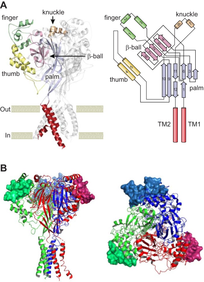
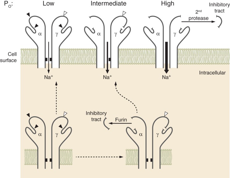
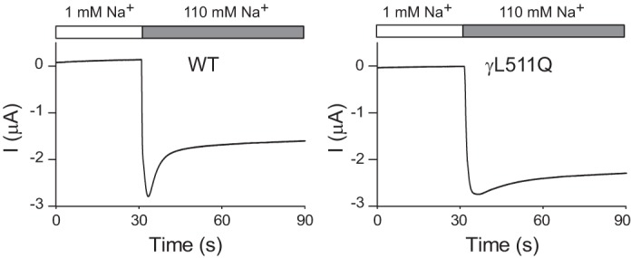
Epithelial Na+ channels (ENaCs) are members of a family of cation channels that function as sensors of the extracellular environment. ENaCs are activated by specific proteases in the biosynthetic pathway and at the cell surface and remove embedded inhibitory tracts, which allows channels to transition to higher open-probability states. Resolved structures of ENaC and an acid-sensing ion channel revealed highly organized extracellular regions. Within the periphery of ENaC subunits are unique domains formed by antiparallel β-strands containing the inhibitory tracts and protease cleavage sites. ENaCs are inhibited by Na+ binding to specific extracellular site(s), which promotes channel transition to a lower open-probability state. Specific inositol phospholipids and channel modification by Cys-palmitoylation enhance channel open probability. How these regulatory factors interact in a concerted manner to influence channel open probability is an important question that has not been resolved. These various factors are reviewed, and the impact of specific factors on human disorders is discussed.

Keywords: ASIC; ENaC; gating; palmitoylation; phosphatidylinositol; protease; sodium.


Conflict of interest statement

T. R. Kleyman receives an honorarium from Wiley, Inc., as Editor-in-Chief of Physiological Reports. D. C. Eaton has no conflicts of interest, financial or otherwise, to disclose.

Figures

Fig. 1.
Fig. 1.
Acid-sensing ion channel (ASIC) type 1 and epithelial Na+ channel (ENaC) structures. A, left: ribbon illustration of the ASIC1 homotrimer, highlighting the extracellular and transmembrane regions of 1 subunit. Discrete domains within the extracellular region include the proximal palm and β-ball formed by β-strands and the peripheral α-helical thumb, finger, and knuckle domains. Transmembrane helices are indicated in red. A, right: organization of an ASIC1 subunit. Contiguous peripheral domains with α-helices (cylinders) arise from noncontiguous proximal domains with β-strands (arrows). TM, transmembrane. B: ribbon illustration of a human ENaC heterotrimer. Residues in the gating relief of inhibition by proteolysis (GRIP) domains that are unique to ENaC and contain the α- and γ-subunit-embedded inhibitory tracts are shown as surface rendering. α-Subunit (red), β-subunit (blue), and γ-subunit (green) are shown. Side (left) and top (right) views are shown. ASIC1 and ENaC structures are based on Protein Data Bank codes 4NYK and 6BQN (75, 121). [Top right and top left were modified from Kashlan and Kleyman (82), with permission.]
Fig. 2.
Fig. 2.
Epithelial Na+ channel (ENaC) activation by proteases. ENaC subunits assemble in the endoplasmic reticulum. As channels transit through the biosynthetic pathway, the α- and γ-subunits are processed by furin, releasing the α-subunit inhibitory tract. This favors transition of channels from a low to an intermediate open probability (Po). The γ-subunit is cleaved once by furin. Cleavage of this subunit by a second protease results in release of its inhibitory tract and favors transition of channels to a high open probability. There are other important factors that influence channel open probability. [Modified from Ray et al. (139), with permission from Elsevier.]
Fig. 3.
Fig. 3.
Epithelial Na+ channel (ENaC) Na+ self-inhibition response. Current traces illustrate the Na+ self-inhibition response assessed in Xenopus oocytes expressing wild-type (WT) human ENaC (left) or the γL511Q mutant (right). Oocytes were initially bathed in a solution containing 1 mM Na and 109 mM N-methyl-d-glucamine. After a rapid transition to a 110 mM Na bath, a rapid increase in inward Na+ current (I; downward deflection) was followed by a slow reduction in current, reflecting Na+ self-inhibition. The Na+ self-inhibition response in oocytes expressing the γL511Q mutant was markedly dampened. [Modified from Chen et al. (38), with permission.]
Fig. 4.
Fig. 4.
Schematic diagram of myristoylated alanine-rich C-kinase substrate (MARCKS) and MARCKS-like protein 1 (MLP-1). The effector domain has multiple sites for phosphatidylinositol 4,5-bisphosphate (PIP2) interaction (note positive-charged residues) and sites for phosphorylation (note serine residues). MARCKS and MLP-1 function as a reversible source of PIP2 at the membrane. MLP-1 is the predominant MARCKS isoform in the mouse kidney. The ability of MARCKS and MLP-1 to function as a PIP2-sequestering protein at the membrane is dependent on hydrophobic interactions between the myristoylation domain and the membrane, and electrostatic forces between the effector domain and anionic lipids in the membrane.
Fig. 5.
Fig. 5.
Interaction of myristoylated alanine-rich C-kinase substrate (MARCKS) with the plasma membrane. Dephosphorylated MARCKS binds to the plasma membrane and cross-links to actin. Phosphorylated MARCKS detaches from the plasma membrane and disrupts actin filaments. PIP2, phosphatidylinositol 4,5-bisphosphate.

References

    1. Abriel H, Loffing J, Rebhun JF, Pratt JH, Schild L, Horisberger JD, Rotin D, Staub O. Defective regulation of the epithelial Na+ channel by Nedd4 in Liddle’s syndrome. J Clin Invest 103: 667–673, 1999. doi:10.1172/JCI5713. - DOI - PMC - PubMed
    1. Adebamiro A, Cheng Y, Rao US, Danahay H, Bridges RJ. A segment of γ-ENaC mediates elastase activation of Na+ transport. J Gen Physiol 130: 611–629, 2007. doi:10.1085/jgp.200709781. - DOI - PMC - PubMed
    1. Alli AA, Bao HF, Alli AA, Aldrugh Y, Song JZ, Ma HP, Yu L, Al-Khalili O, Eaton DC. Phosphatidylinositol phosphate-dependent regulation of Xenopus ENaC by MARCKS protein. Am J Physiol Renal Physiol 303: F800–F811, 2012. doi:10.1152/ajprenal.00703.2011. - DOI - PMC - PubMed
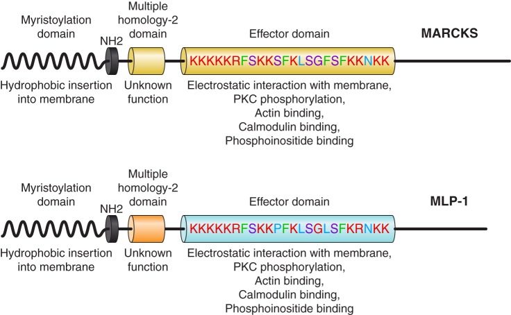
    1. Alli AA, Bao HF, Liu BC, Yu L, Aldrugh S, Montgomery DS, Ma HP, Eaton DC. Calmodulin and CaMKII modulate ENaC activity by regulating the association of MARCKS and the cytoskeleton with the apical membrane. Am J Physiol Renal Physiol 309: F456–F463, 2015. doi:10.1152/ajprenal.00631.2014. - DOI - PMC - PubMed
    1. Alli AA, Song JZ, Al-Khalili O, Bao HF, Ma HP, Alli AA, Eaton DC. Cathepsin B is secreted apically from Xenopus 2F3 cells and cleaves the epithelial sodium channel (ENaC) to increase its activity. J Biol Chem 287: 30073–30083, 2012. doi:10.1074/jbc.M111.338574. - DOI - PMC - PubMed

Publication types

LinkOut - more resources