. 2019 Nov 13;14(11):e0225025.
doi: 10.1371/journal.pone.0225025. eCollection 2019.

Equine bronchial fibroblasts enhance proliferation and differentiation of primary equine bronchial epithelial cells co-cultured under air-liquid interface

Affiliations

Equine bronchial fibroblasts enhance proliferation and differentiation of primary equine bronchial epithelial cells co-cultured under air-liquid interface

Vanessa Abs et al. PLoS One. .

Abstract

Interaction between epithelial cells and fibroblasts play a key role in wound repair and remodelling in the asthmatic airway epithelium. We present the establishment of a co-culture model using primary equine bronchial epithelial cells (EBECs) and equine bronchial fibroblasts (EBFs). EBFs at passage between 4 and 8 were seeded on the bottom of 24-well plates and treated with mitomycin C at 80% confluency. Then, freshly isolated (P0) or passaged (P1) EBECs were seeded on the upper surface of membrane inserts that had been placed inside the EBF-containing well plates and grown first under liquid-liquid interface (LLI) then under air-liquid interface (ALI) conditions to induce epithelial differentiation. Morphological, structural and functional markers were monitored in co-cultured P0 and P1 EBEC monolayers by phase-contrast microscopy, scanning and transmission electron microscopy, hematoxylin-eosin, immunocytochemistry as well as by measuring the transepithelial electrical resistance (TEER) and transepithelial transport of selected drugs. After about 15-20 days of co-culture at ALI, P0 and P1 EBEC monolayers showed pseudo-stratified architecture, presence of ciliated cells, typically honeycomb-like pattern of tight junction protein 1 (TJP1) expression, and intact selective barrier functions. Interestingly, some notable differences were observed in the behaviour of co-cultured EBECs (adhesion to culture support, growth rate, differentiation rate) as compared to our previously described EBEC mono-culture system, suggesting that cross-talk between epithelial cells and fibroblasts actually takes place in our current co-culture setup through paracrine signalling. The EBEC-EBF co-culture model described herein will offer the opportunity to investigate epithelial-mesenchymal cell interactions and underlying disease mechanisms in the equine airways, thereby leading to a better understanding of their relevance to pathophysiology and treatment of equine and human asthma.


Conflict of interest statement

The authors have declared that no competing interests exist.

Figures

Fig 1
Fig 1. Schematic representation of the EBEC-EBF transwell co-culture model under liquid-liquid (LLI) and air-liquid interface (ALI) condition.
Equine bronchial fibroblasts (EBFs) at passages between 4 and 8 were seeded on 24-well plates and treated with mitomyin C when they reached 80% confluency. Freshly isolated (P0) or passaged (P1) EBECs were seeded on membrane inserts that were placed inside the EBF-containing well plates. EBECs were first allowed to grow under liquid-liquid interface (LLI) conditions (with complete DMEM in the basal compartment and complete AECGM in the apical compartment) until they formed confluent monolayers, then air-liquid interface (ALI) conditions (with ALI medium in the basal compartment and air in the apical compartment) were established to induce epithelial differentiation.
Fig 2
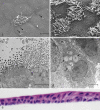
Fig 2. Phenotypic features of morphological, structural and ultrastructural differentiation detected in P0 and P1 EBEC monolayers during co-culture with mitomycin C-treated EBFs under ALI-conditions.
Representative SEM micrographs revealing the presence of cilia after 8 days (a) and 20 days (b) of co-culture at ALI, as well as of microvilli (a, b) and mucus-like amorphous material (a) (arrows). TEM micrographs showing cross sections of a number of cilia (c) with typical internal structures of their basal body or centriole (the so called 9+0 arrangement of microtubules, consisting of a circle of 9 sets of triplets embedded in the apical part of the epithelial cells; black arrow heads) and motile part (the so-called 9+2 arrangement of microtubules, consisting of a circle of 9 doublets and one central doublet; black arrows), cross sections of a number of microvilli and a mucus-containing cell (d, black arrows). Representative images of H&E- (e) stained sections of “matured” epithelial layers (15–20 days of ALI) showing pseudo-stratified columnar architecture (e; magnification: x40). The scale bar for (a) was 10 μM, for (b) 5 μM as well as for both c and d 2 μM, respectively.
Fig 3
Fig 3. Purity of EBEC and EBF monolayers in co-culture under ALI conditions as depicted in Fig 1.
Representative phase-contrast microscopy images [magnification: x4] showing the morphological appearance of “mature” confluent P1 EBEC monolayers after 15–20 days of co-culture at ALI (a; P1) and of 80% confluent monolayers of mitomycin C-treated EBFs maintained in ALI-medium for the same culture period (d; P6)). Representative images of immunofluorescence staining of confluent P1 EBEC monolayers after 15–20 days of co-culture at ALI for the epithelial cell marker cytokeratin (b P0; and c nuclei blue stained with DAPI) and the mesenchymal cell marker vimentin (rest of green fluorescence in EBEC P0 c; e) [magnification: x40; green fluorescence (FITC) is positive signal; nuclei are blue stained with DAPI].
Fig 4
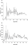
Fig 4. Bioelectrical characteristics of EBECs grown on insert membranes in co-culture with mitomycin C-treated EBFs.
The graphs show the time-course of TEER development in P0 (a) and P1 (b) co-cultured EBEC monolayers. The abscissa indicates co-culture days before (negative) and after (positive) switch to the ALI condition (day 0). Each point represents mean ± SEM (n = 5, except for the TEER graph of P1 EBECs from ALI day 20 to ALI day 60, where n = 3).
Fig 5
Fig 5. Time-course of tight junction expression pattern in EBEC monolayers grown in the presence of monolayers of mitomycin C-treated EBFs.
Representative images of confluent P0 (a-c) and P1 (d-f) EBEC monolayers immunolabelled for TJP1 (green fluorescence) immediately before switching from the LLI to the ALI condition (ALI day 0) (a, d), shortly after the achievement of a stable TEER value under ALI conditions (P0: ALI days 10–15; P1: ALI days 7–10 days) (b, e) and in more “mature” co-cultures (ALI days 15–20) (c, f). Magnification: x40; nuclei are blue stained with DAPI.
Fig 6
Fig 6. The apparent permeability coefficients (Papp) for the apical-to-basolateral transport of atenolol and propranolol across P0 EBEC monolayers co-cultured under ALI conditions with mitomycin C-treated EBFs.
The graph compares the mean (± SEM) permeability values (Papp) calculated for the paracellular transport marker atenolol and the transcellular transport marker propranolol with time in co-culture P0 EBEC monolayers at ALI for 7, 9 and 11 days (n = 5) and across blank (cell-free) inserts (n = 5) after 10 (a), 30 (b) and 50 (c) min of incubation. Data are depicted as means ± S.E.M from n = 5–8 independent experiments. Asterisks indicate statistical significance from blank values and between drugs; *(p<0.05), **(p<0.01), ***(p<0.001) and ns: not significant.

References

    1. Holgate ST, Lackie PM, Davies DE, Roche WR, Walls AF. The bronchial epithelium as a key regulator of airway inflammation and remodelling in asthma. Clin Exp Allergy 1999; 29 Suppl. 2: 90–95. - PubMed
    1. Xiao C, Puddicombe SM, Field S, Haywood J, Broughton-Head V, Puxeddu I, et al. Defective epithelial barrier function in asthma. J Allergy Clin Immunol. 2011; 128: 549–556. 10.1016/j.jaci.2011.05.038 - DOI - PubMed
    1. Humlicek AL, Manzel LJ, Chin CL, Shi L, Excoffon KJ, Winter MC, et al. Paracellular permeability restricts airway epithelial responses to selectively allow activation by mediators at the basolateral surface. J Immunol. 2007; 178: 6395–6403. 10.4049/jimmunol.178.10.6395 - DOI - PubMed
    1. Coyne CB, Vanhook MK, Gambling TM, Carson JL, Boucher RC, Johnson LG. Regulation of airway tight junctions by proinflammatory cytokines. Mol Biol Cell. 2002; 13: 3218–3234. 10.1091/mbc.E02-03-0134 - DOI - PMC - PubMed
    1. Haag S, Matthiesen S, Juergens UR, Racké K. Muscarinic receptors mediate stimulation of collagen synthesis in human lung fibroblasts. Eur Respir J. 2008; 32: 555–562. 10.1183/09031936.00129307 - DOI - PubMed

Publication types

MeSH terms