Review
. 2020 May;77(10):1933-1945.
doi: 10.1007/s00018-019-03373-1. Epub 2019 Nov 13.

Vertebrate cranial mesoderm: developmental trajectory and evolutionary origin

Affiliations
Review

Vertebrate cranial mesoderm: developmental trajectory and evolutionary origin

Bhakti Vyas et al. Cell Mol Life Sci. 2020 May.

Abstract

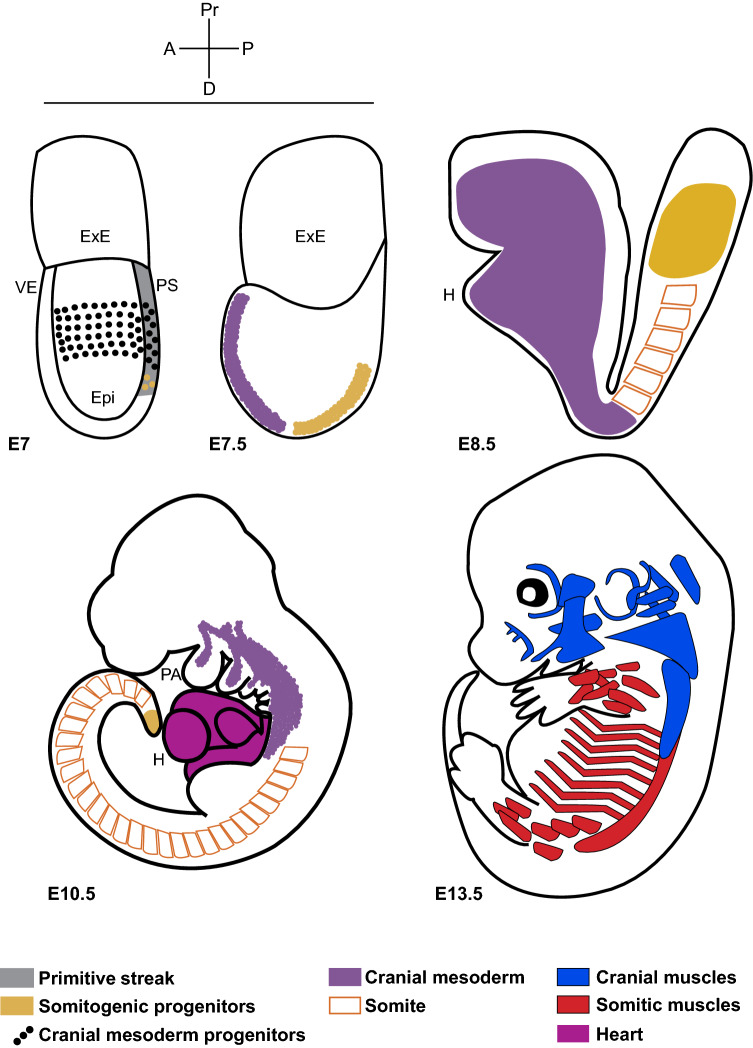
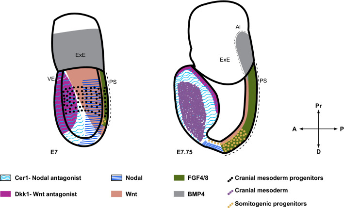
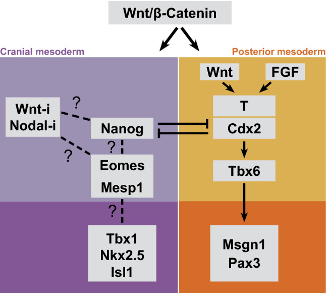
Vertebrate cranial mesoderm is a discrete developmental unit compared to the mesoderm below the developing neck. An extraordinary feature of the cranial mesoderm is that it includes a common progenitor pool contributing to the chambered heart and the craniofacial skeletal muscles. This striking developmental potential and the excitement it generated led to advances in our understanding of cranial mesoderm developmental mechanism. Remarkably, recent findings have begun to unravel the origin of its distinct developmental characteristics. Here, we take a detailed view of the ontogenetic trajectory of cranial mesoderm and its regulatory network. Based on the emerging evidence, we propose that cranial and posterior mesoderm diverge at the earliest step of the process that patterns the mesoderm germ layer along the anterior-posterior body axis. Further, we discuss the latest evidence and their impact on our current understanding of the evolutionary origin of cranial mesoderm. Overall, the review highlights the findings from contemporary research, which lays the foundation to probe the molecular basis of unique developmental potential and evolutionary origin of cranial mesoderm.

Keywords: Cardiopharyngeal field; Head mesoderm; Head muscles; Mesoderm development; Vertebrate head evolution.


Figures

Fig. 1
Fig. 1
Schematic illustrating the origin and derivatives of cranial mesoderm. Cartoons of mouse embryos from mid-gastrula stage showing cranial and somitic mesoderm progenitors and their muscle derivatives. Embryonic day E7 all mesodermal progenitors emerge from the posterior end, where the primitive streak is formed. E7 and E7.5 cranial mesoderm migrates to the anterior pole, while somitogenic progenitors develop on the posterior side. E8.5 and E10.5 lateral plate of cranial mesoderm forms the heart and the paraxial component spreads as a non-segmented mesenchyme and eventually patterns into streams entering the pharyngeal arches. This contrasts with posterior paraxial mesoderm forming somites. E13.5 the pharyngeal mesodermal core contributes to different cranial skeletal muscle groups (in blue). Somite-derived skeletal muscles are shown in red. VE visceral endoderm, ExE extra-embryonic ectoderm, Epi epiblast, PS primitive streak (marked in grey), H heart, PA pharyngeal arches, A anterior, P posterior, Pr proximal, D distal. E7, 7.5 (based on evidence from [40]), E8.5 (based on evidence from Mesp1-cre/R26R; [153]), E10.5 [129] and E13.5 [53]
Fig. 2
Fig. 2
Signaling cues driving progressive specification of cranial and posterior somitogenic mesoderm. Schematic highlighting the expression domains of key signaling cues influencing mesoderm patterning. Anatomical locations of cranial and somitic mesoderm and their progenitors at these temporal windows are indicated. A anterior, P posterior, Pr proximal, D distal, ExE extra-embryonic ectoderm, Al Allantois (marked by dotted line), VE visceral endoderm, PS primitive streak (marked by dashed line)
Fig. 3
Fig. 3
Divergent regulatory network governing cranial and posterior somitogenic mesoderm. Wnt/β-catenin signal is central for the generation of all mesoderm. The regulatory network of somite-forming posterior mesoderm is well studied. Recent studies reveal key components of the cranial mesoderm program; however, the network is unclear. Wnt-i Wnt inhibition, Nodal-i Nodal inhibition

References

    1. Gans C, Northcutt RG. Neural crest and the origin of vertebrates: a new head. Science (80-) 1983;220:268–273. doi: 10.1126/science.220.4594.268. - DOI - PubMed
    1. Stolfi A, et al. Early chordate origins of the vertebrate second heart field. Science (80-) 2010 doi: 10.1126/science.1190181. - DOI - PMC - PubMed
    1. Kaplan N, Razy-Krajka F, Christiaen L. Regulation and evolution of cardiopharyngeal cell identity and behavior: insights from simple chordates. Curr Opin Genet Dev. 2015;32:119–128. doi: 10.1016/j.gde.2015.02.008. - DOI - PMC - PubMed
    1. Delsuc F, et al. Tunicates and not cephalochordates are the closest living relatives of vertebrates. Nature. 2006;439:965–968. doi: 10.1038/nature04336. - DOI - PubMed
    1. Sambasivan R, Kuratani S, Tajbakhsh S. An eye on the head: the development and evolution of craniofacial muscles. Development. 2011;138:2401–2415. doi: 10.1242/dev.040972. - DOI - PubMed

MeSH terms

LinkOut - more resources