. 2019 Nov 12;29(7):1946-1960.e5.
doi: 10.1016/j.celrep.2019.10.029.

Insulin Potentiates JAK/STAT Signaling to Broadly Inhibit Flavivirus Replication in Insect Vectors

Affiliations

Insulin Potentiates JAK/STAT Signaling to Broadly Inhibit Flavivirus Replication in Insect Vectors

Laura R H Ahlers et al. Cell Rep. .

Abstract

The World Health Organization estimates that more than half of the world's population is at risk for vector-borne diseases, including arboviruses. Because many arboviruses are mosquito borne, investigation of the insect immune response will help identify targets to reduce the spread of arboviruses. Here, we use a genetic screening approach to identify an insulin-like receptor as a component of the immune response to arboviral infection. We determine that vertebrate insulin reduces West Nile virus (WNV) replication in Drosophila melanogaster as well as WNV, Zika, and dengue virus titers in mosquito cells. Mechanistically, we show that insulin signaling activates the JAK/STAT, but not RNAi, pathway via ERK to control infection in Drosophila cells and Culex mosquitoes through an integrated immune response. Finally, we validate that insulin priming of adult female Culex mosquitoes through a blood meal reduces WNV infection, demonstrating an essential role for insulin signaling in insect antiviral responses to human pathogens.

Keywords: Culex quinquefasciatus; DGRP; Drosophila melanogaster; ERK; Kunjin virus; West Nile virus; Zika virus; dengue virus; innate immunity; mosquito.


Conflict of interest statement

DECLARATION OF INTERESTS

A.G.G., S.L., L.R.H.A., and C.E.T. have filed a provisional patent application (62/810754) related to the use of insulin receptor agonists.

Figures

Figure 1.
Figure 1.. A Genetic Screen of D. melanogaster Identified Candidate Genes Involved in the D. melanogaster Response to WNV-Kun
(A) Schematic of the screen and downstream analysis. (B) Survival of each DGRP line, measured by log(hazard ratio). (C) Gene set enrichment analysis (GSEA) using genes for all variants and their associated p values from the GWAS. Heatmap data represent GWAS variant p values, while the bar graph indicates the GSEA p value and the number of genes enriched for each GO category in parentheses. Genes indicated in orange boxes are components of the JAK/STAT pathway, and genes indicated by blue boxes are components of the insulin signaling pathway.
Figure 2.
Figure 2.. Components of the JAK/STAT and Insulin Signaling Pathways Are Necessary for D. melanogaster Survival against WNV-Kun Infection
(A–C) Mutants in the genes (B) upd2 and upd3 and (C) hop are susceptible to WNV-Kun infection compared with the (A) y1w1 isotype control. (D) WNV-Kun titer is higher in Stat92E and vir-1 mutant flies relative to the y1w1 isotype control at 5 days post-infection (#p < 0.05, Mann-Whitney test). (E) InR knockdown and (F) ilp71 mutant flies are susceptible to WNV-Kun compared with the sibling or background controls. Hazard ratio for each infection group is indicated in parenthesis, and statistical significance from the control group is indicated with an asterisk (*p < 0.05, log-rank test). Each survival curve represents two (B and C) or three (A, E, and F) independent experiments of >40 flies that were combined for a final survival curve. For titer results (D), marker shapes represent biological replicates, the bar represents the mean, and error bars represent SDs. Samples for which virus was not detected are indicated by “n.d.” Titer data are representative of duplicate independent experiments.
Figure 3.
Figure 3.. The D. melanogaster Insulin Signaling Pathway Is Activated by Vertebrate Insulin, Which Is Antiviral to WNV-Kun
(A) Akt is phosphorylated and activated in D. melanogaster S2 cells when treated with bovine insulin. (B) WNV-Kun titer is reduced in S2 cells primed with 1.7 μM bovine insulin for 24 h prior to infection (MOI = 0.01 PFU/cell). (C) WNV-Kun titer is reduced in D. melanogaster on a diet of 10 μM bovine insulin (*p < 0.05, unpaired t test). Open circles represent biological replicates. Horizontal lines or bars represent the mean. Error bars represent SDs. (D) Ingested insulin disseminates from the midgut into the hemolymph in flies on a diet of 10 μM bovine insulin. Results are representative of duplicate (A and D) or triplicate (B and C) independent experiments.
Figure 4.
Figure 4.. Insulin Priming Activates Antiviral Pathways in D. melanogaster
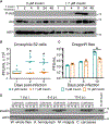
(A–G) Induction of genes within the JAK/STAT pathway, (A) upd2, (B) upd3, (C) TotM, and (D) vir-1, and the RNAi pathway, (E) AGO1, (F) AGO2, and (G) Dcr2, were measured using qRT-PCR following priming of D. melanogaster S2 cells with 1.7 μM insulin and mock or WNV-Kun infection. (H) Levels of ERK, Akt, and Stat92E phosphorylation were measured using western blot following insulin treatment and mock or WNV-Kun infection of S2 cells. (I) FoxO localization in the larval fat body was determined by confocal microscopy using third-instar larvae on a standard cornmeal diet with or without 10 μM bovine insulin and mock- or WNV-Kun-infected for 4 h. (J–M) ERK was knocked down in S2 cells (J), and transcript levels of (K) upd2, (L) upd3, and (M) vir-1 were measured using qRT-PCR (*p < 0.05, **p < 0.01, ***p < 0.001, and ****p < 0.0001, ANOVA with correction for multiple comparisons). Open circles represent biological replicates. Horizontal black bars represent the mean. Error bars represent SDs. Results are representative of duplicate independent experiments.
Figure 5.
Figure 5.. Insulin Priming Reduces Flavivirus Titer in Cx. quinquefasciatus, Ae. aegypti, and Ae. albopictus Cells
(A–C) Akt is phosphorylated and activated by insulin priming in (A) Cx. quinquefasciatus Hsu cells, (B) Ae. aegypti Aag2 cells, and (C) Ae. albopictus C6/36 cells. (D–H) Insulin priming reduces WNV-Kun titer in (D) Hsu, (E) Aag2, and (F) C6/36 cells and (G) Zika virus and (H) dengue virus titer in C6/36 cells (MOI = 0.01 PFU/cell) (*p < 0.05, **p < 0.01, ***p < 0.001, ****p < 0.0001, unpaired t test). Open circles represent biological replicates. Horizontal bars represent the mean. Error bars represent SDs. Results are representative of triplicate independent experiments.
Figure 6.
Figure 6.. Insulin Activates the Antiviral Response in Adult Female Cx. quinquefasciatus
(A) Schematic illustrating process of age-matching pupae into adults, feeding female mosquitoes a blood meal of chicken blood with or without insulin or WNV-Kun, and collecting adults for analysis post-infection. (B–F) Induction of (B) CxR2D2, (C) CxDcr2, (D) CxSTAT, (E) WNV-Kun envelope (env), and (F) NS5 genes were measured using qRT-PCR following blood feeding of Cx. quinquefasciatus (*p < 0.05, unpaired t test; or #p < 0.05, Mann-Whitney test). Circles represent biological replicates. Bars represent the mean. Error bars represent SDs. Results are representative of duplicate independent experiments.
Figure 7.
Figure 7.. Schematic of Immune Signaling during Insulin Priming in Insects
Insulin (red box) binds to the insulin-like receptor (InR), activating a signaling cascade that inhibits FoxO-dependent transcription of RNAi components and reduces RNAi-dependent antiviral immunity. Proposed networked regulation of insulin and JAK/STAT signaling includes activation of ERK downstream of PI3K and increased expression of upd2/3, suggesting a control point for insulin-enhanced JAK/STAT signaling. Signaling components denoted by pink stars were important for D. melanogaster survival during WNV-Kun infection. Components indicated by orange boxes were induced at the transcript level by insulin treatment, and those indicated by teal boxes were activated at the protein level.

References

    1. Ahlers LRH, Bastos RG, Hiroyasu A, and Goodman AG (2016). Invertebrate iridescent virus 6, a DNA virus, stimulates a mammalian innate immune response through RIG-I-like receptors. PLoS ONE 11, e0166088. - PMC - PubMed
    1. Arbouzova NI, and Zeidler MP (2006). JAK/STAT signalling in Drosophila: insights into conserved regulatory and cellular functions. Development 133, 2605–2616. - PubMed
    1. Arensburger P, Megy K, Waterhouse RM, Abrudan J, Amedeo P, Antelo B, Bartholomay L, Bidwell S, Caler E, Camara F, et al. (2010). Sequencing of Culex quinquefasciatus establishes a platform for mosquito comparative genomics. Science 330, 86–88. - PMC - PubMed
    1. Arik AJ, Hun LV, Quicke K, Piatt M, Ziegler R, Scaraffia PY, Badgandi H, and Riehle MA (2015). Increased Akt signaling in the mosquito fat body increases adult survivorship. FASEB J. 29, 1404–1413. - PMC - PubMed
    1. Aytug S, Reich D, Sapiro LE, Bernstein D, and Begum N (2003). Impaired IRS-1/PI3-kinase signaling in patients with HCV: a mechanism for increased prevalence of type 2 diabetes. Hepatology 38, 1384–1392. - PubMed

Publication types

LinkOut - more resources