. 2019 Dec 16;47(22):11746-11754.
doi: 10.1093/nar/gkz978.

G-quadruplex structures trigger RNA phase separation

Affiliations

G-quadruplex structures trigger RNA phase separation

Yueying Zhang et al. Nucleic Acids Res. .

Abstract

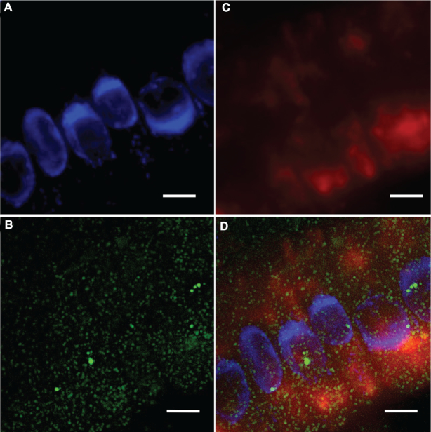
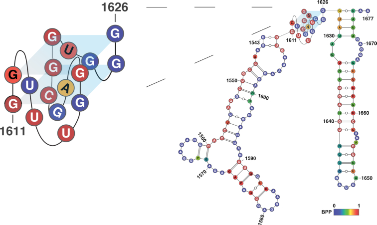
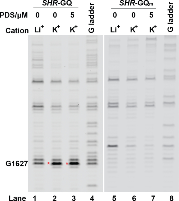
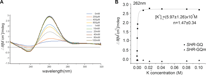
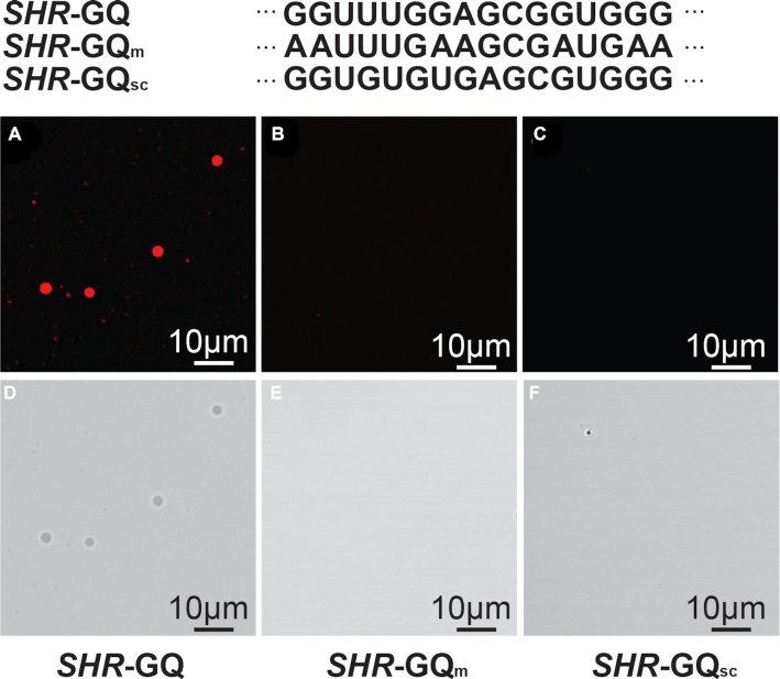
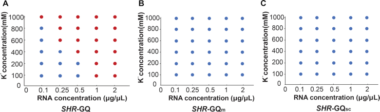
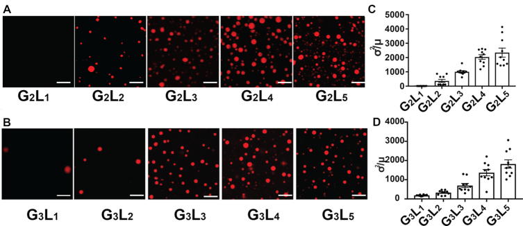
Liquid-liquid phase separation plays an important role in a variety of cellular processes, including the formation of membrane-less organelles, the cytoskeleton, signalling complexes, and many other biological supramolecular assemblies. Studies on the molecular basis of phase separation in cells have focused on protein-driven phase separation. In contrast, there is limited understanding on how RNA specifically contributes to phase separation. Here, we described a phase-separation-like phenomenon that SHORT ROOT (SHR) RNA undergoes in cells. We found that an RNA G-quadruplex (GQ) forms in SHR mRNA and is capable of triggering RNA phase separation under physiological conditions, suggesting that GQs might be responsible for the formation of the SHR phase-separation-like phenomenon in vivo. We also found the extent of GQ-triggered-phase-separation increases on exposure to conditions which promote GQ. Furthermore, GQs with more G-quartets and longer loops are more likely to form phase separation. Our studies provide the first evidence that RNA can adopt structural motifs to trigger and/or maintain the specificity of RNA-driven phase separation.


Figures

Figure 1.
Figure 1.
Phase-separation-like SHR and punctate SCR RNA labelling observed in plant root cells, images generated by smFISH. (A) Nuclei stained with DAPI (blue), SCR RNA (green) is shown in (B) and SHR RNA (red) in (C). Merged image in (D). Scale bar, 10 μm.
Figure 2.
Figure 2.
Formation of GQ in SHR mRNA. RNA structure model of SHR, showing the fragment with GQ structure, predicted by ViennaRNA (24). A parallel topology is shown (supported by circular dichroism studies), nucleotides are color-coded according to base-pairing probability (BPP), where 1 represent 100% double-stranded, 0 represent 100% single-stranded. GQ structure is enlarged for better visualization.
Figure 3.
Figure 3.
Reverse transcription stalling assays of SHR-GQ RNA shows K+ dependent stalling (red asterisk) at one nucleotide after the folded GQ (lane 2 and lane 3), SHR-GQm do not have stalling at the corresponding position (lane 6 and lane 7). G-ladders (lane 4 and lane 8) show the sequencing lanes of G for SHR-GQ (lane 4) and SHR-GQm (lane 8), respectively.
Figure 4.
Figure 4.
SHR-GQ forms a parallel GQ structure. (A) Circular Dichroism profile of SHR-GQ RNA with potassium ion titration. (B) Circular Dichroism signal (ellipticity monitored at 262 nm) of SHR-GQ (squares) and SHR-GQm (triangles) are shown as a function of K+ concentration. Data were fitted with Hill equation. The [K+]1/2 and Hill coefficients (n) are provided in the plot.
Figure 5.
Figure 5.
GQ is able to trigger the liquid–liquid phase separation. Fluorescence micrographs of SHR-GQ (AD), GQ-mutation (SHR-GQm) (BE) and GQ-scramble (SHR-GQsc) (CF) RNAs at the physiological K+ concentration. Scale bars = 10 μm
Figure 6.
Figure 6.
Phase diagrams of SHR-GQ (A), SHR-GQm (B) and SHR-GQsc (C) under different RNA and K+ concentrations. Red dots indicate where liquid–liquid-phase-separation occurs; blue dots indicate a lack of liquid–liquid-phase-separation.
Figure 7.
Figure 7.
The quick rearrangement of GQ-triggered droplets. Left, droplets formed by SHR-GQ. Right, droplets formed by SHR-GQ undergo fusion over time. Scale bar = 5 μm.
Figure 8.
Figure 8.
The capability for GQ-triggered-phase-separation is affected by the number of G-quartets and loop lengths. (A) Fluorescence micrographs of phase separation of G2 GQ RNAs with different loop lengths (1 μg/μl RNA under 800mM K+). (B) Fluorescence micrographs of phase separation of G3 GQ RNAs with different loop lengths (1 μg/μl RNA under 400 mM K+). (C) Quantitation of inhomogeneity as normalized variance (σ2/μ) of phase separation for G2 GQ RNAs with different loop lengths. (D) Quantitation of inhomogeneity as normalized variance (σ2/μ) of phase separation for G3 GQ RNAs with different loop lengths. Data are shown as mean ± SE.

References

    1. Hyman A.A., Weber C.A., Jülicher F.. Liquid-liquid phase separation in biology. Annu. Rev. Cell Dev. Biol. 2014; 30:39–58. - PubMed
    1. Brangwynne C.P., Eckmann C.R., Courson D.S., Rybarska A., Hoege C., Gharakhani J., Jülicher F., Hyman A.A.. Germline p granules are liquid droplets that localize by controlled dissolution/condensation. Science. 2009; 324:1729. - PubMed
    1. Brangwynne C.P., Mitchison T.J., Hyman A.A.. Active liquid-like behavior of nucleoli determines their size and shape in Xenopus laevis oocytes. Proc. Natl. Acad. Sci. U.S.A. 2011; 108:4334. - PMC - PubMed
    1. Woodruff J.B., Ferreira Gomes B., Widlund P.O., Mahamid J., Honigmann A., Hyman A.A.. The centrosome is a selective condensate that nucleates microtubules by concentrating tubulin. Cell. 2017; 169:1066–1077. - PubMed
    1. Hernández-Vega A., Braun M., Scharrel L., Jahnel M., Wegmann S., Hyman B.T., Alberti S., Diez S., Hyman A.A.. Local nucleation of microtubule bundles through tubulin concentration into a condensed tau phase. Cell Rep. 2017; 20:2304–2312. - PMC - PubMed

Publication types

LinkOut - more resources