. 2019 Nov 13;4(6):e00536-19.
doi: 10.1128/mSphere.00536-19.

A Metabolic Dependency for Host Isoprenoids in the Obligate Intracellular Pathogen Rickettsia parkeri Underlies a Sensitivity to the Statin Class of Host-Targeted Therapeutics

Affiliations

A Metabolic Dependency for Host Isoprenoids in the Obligate Intracellular Pathogen Rickettsia parkeri Underlies a Sensitivity to the Statin Class of Host-Targeted Therapeutics

Vida Ahyong et al. mSphere. .

Erratum in

Abstract

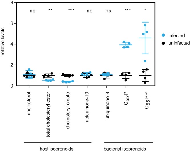
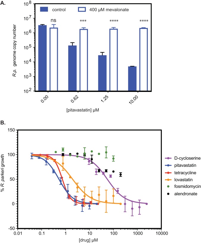
Gram-negative bacteria in the order Rickettsiales have an obligate intracellular growth requirement, and some species cause human diseases such as typhus and spotted fever. The bacteria have evolved a dependence on essential nutrients and metabolites from the host cell as a consequence of extensive genome reduction. However, it remains largely unknown which nutrients they acquire and whether their metabolic dependency can be exploited therapeutically. Here, we describe a genetic rewiring of bacterial isoprenoid biosynthetic pathways in the Rickettsiales that has resulted from reductive genome evolution. Furthermore, we investigated whether the spotted fever group Rickettsia species Rickettsia parkeri scavenges isoprenoid precursors directly from the host. Using targeted mass spectrometry, we found that infection caused decreases in host isoprenoid products and concomitant increases in bacterial isoprenoid metabolites. Additionally, we report that treatment of infected cells with statins, which inhibit host isoprenoid synthesis, prohibited bacterial growth. We show that growth inhibition correlates with changes in bacterial size and shape that mimic those caused by antibiotics that inhibit peptidoglycan biosynthesis, suggesting that statins lead to an inhibition of cell wall synthesis. Altogether, our results describe a potential Achilles' heel of obligate intracellular pathogens that can potentially be exploited with host-targeted therapeutics that interfere with metabolic pathways required for bacterial growth.IMPORTANCE Obligate intracellular pathogens, which include viruses as well as certain bacteria and eukaryotes, are a subset of infectious microbes that are metabolically dependent on and unable to grow outside an infected host cell because they have lost or lack essential biosynthetic pathways. In this study, we describe a metabolic dependency of the bacterial pathogen Rickettsia parkeri on host isoprenoid molecules that are used in the biosynthesis of downstream products, including cholesterol, steroid hormones, and heme. Bacteria make products from isoprenoids, such as an essential lipid carrier for making the bacterial cell wall. We show that bacterial metabolic dependency can represent a potential Achilles' heel and that inhibiting host isoprenoid biosynthesis with the FDA-approved statin class of drugs inhibits bacterial growth by interfering with the integrity of the cell wall. This work supports the potential to treat infections by obligate intracellular pathogens through inhibition of host biosynthetic pathways that are susceptible to parasitism.

Keywords: Rickettsia; isoprenoids; metabolic parasitism.


Figures

FIG 1
FIG 1
Generalized MEV and MEP pathways leading to downstream isoprenoid products. The green arrows indicate the presence of annotated enzymes encoded in the R. parkeri genome. The gray arrows indicate the absence of annotated enzymes encoded in the R. parkeri genome. The orange arrow indicates the presence of the IDI enzyme from the host genome. The yellow background denotes the upstream isoprenoid pathway, whereas the blue denotes the downstream isoprenoid pathway. The central black arrow and white question mark denote the possible presence of transporters for isoprenoids from the host into the bacteria. Arrows with double hash marks indicate multiple enzymatic steps. Statin drugs inhibit the activity of HMG-CoA reductase and the formation of mevalonate of the MEV pathway. Fosmidomycin is an inhibitor of DXP reductoisomerase of the MEP pathway. Alendronate sodium hydrate is an inhibitor of farnesyl diphosphate synthase. d-Cycloserine (DCS) is an inhibitor of d-alanine racemase and d-alanine-d-alanine ligase.
FIG 2
FIG 2
Genome evolution in the Rickettsiales isoprenoid pathway. (A) Schematic cladogram of the order Rickettsiales. The presence of intact upstream MEP pathway genes or of the idi gene for each species is indicated. The environmental niche is indicated on the right as follows: C, intracellular cytoplasmic; V, intracellular vacuolar. (B) A schematic of the R. parkeri idi genome locus. Genes involved in conjugal transfer and pseudogenes are indicated. The idi gene is highlighted in purple. (C) Whole-genome alignment of R. parkeri and R. rickettsii. The R. rickettsii genome has a large 33.5-kb deletion (in orange) in the genome alignment for the region adjacent to idi. The gray line segments indicate inverted alignment orientation between R. parkeri and R. rickettsii.
FIG 3
FIG 3
Targeted mass spectrometry of bacterial and host isoprenoids. Shown is a graph of the internal standard equivalent levels of host and bacterial isoprenoids at 4 dpi. Four technical replicates were done. Error bars represent standard deviations. Statistical comparisons were done by an unpaired Student's t test (ns, not significant; **, P < 0.01; ***, P < 0.001; ****, P < 0.0001, for results compared to those with the controls).
FIG 4
FIG 4
Chemical inhibition and rescue of R. parkeri growth. (A) Graph of R. parkeri genome copy numbers in the presence of various concentrations of pitavastatin, without or with mevalonate. Two independent biological replicates were performed, but four technical replicates from a single experiment are shown for simplicity. Error bars represent standard deviations. Statistical comparisons were done by an unpaired Student's t test for each concentration of pitavastatin for the wild type versus the wild type with mevalonate supplementation (ns, not significant; ***, P < 0.001; ****, P < 0.0001). R.p., R. parkeri. (B) Dose-dependent growth inhibition of R. parkeri in the presence of the indicated drugs targeting the MEV or MEP pathway enzymes or of tetracycline, normalized to that of a no-drug control. Fosmidomycin and alendronate failed to generate fit curves.
FIG 5
FIG 5
Shape and size measurements of R. parkeri bacteria under pressure from different drugs. (A) Representative bacterial cells from the EC curve drug concentrations from panel B. Scale bar, 1 μm. (B) Dose-dependent changes in area and eccentricity of R. parkeri bacterial cells under drug treatment with lovastatin, pitavastatin, tetracycline, or d-cycloserine. Each measurement is the mean of at least 40 individual bacterial cells, except for concentrations of d-cycloserine at >250 μM, where most bacterial cells were lysed and fewer measurements were possible (for 500 μM d-cycloserine, n = 22; for 1,000 μM d-cycloserine, n = 18; for 2,000 μM d-cycloserine, n = 13). Error bars represent the 95% confidence interval. (C) Graphs plotting area and eccentricity measurements at the EC100 values calculated from the data shown in Fig. 4B and matched to measurements from panel A. Regions shaded in yellow are the calculated values of EC50 to EC100 from the data shown in Fig. 4B. Graphs show the mean of the sample set, and error bars represent the 95% confidence interval. Statistical comparisons were done by one-way ANOVA of results for all drug-treated samples compared to results for the no-drug (nd) sample set (ns, not significant; **, P < 0.01; ***, P < 0.001; ****, P < 0.0001). dcs, d-cycloserine; tet, tetracycline; lov, lovastatin; pit, pitavastatin.

References

    1. Yu X-J, Walker DH. 2006. The order Rickettsiales, p 493–528. In Dworkin M, Falkow S, Rosenberg E, Schleifer KH, Stackebrandt E (ed), The Prokaryotes. Springer, New York, NY.
    1. Kirkland KB, Wilkinson WE, Sexton DJ. 1995. Therapeutic delay and mortality in cases of Rocky Mountain spotted fever. Clin Infect Dis 20:1118–1121. doi:10.1093/clinids/20.5.1118. - DOI - PubMed
    1. Paddock CD, Sumner JW, Comer JA, Zaki SR, Goldsmith CS, Goddard J, McLellan SLF, Tamminga CL, Ohl CA. 2004. Rickettsia parkeri: a newly recognized cause of spotted fever rickettsiosis in the United States. Clin Infect Dis 38:805–811. doi:10.1086/381894. - DOI - PubMed
    1. Paddock CD, Finley RW, Wright CS, Robinson HN, Schrodt BJ, Lane CC, Ekenna O, Blass MA, Tamminga CL, Ohl CA, McLellan SLF, Goddard J, Holman RC, Openshaw JJ, Sumner JW, Zaki SR, Eremeeva ME. 2008. Rickettsia parkeri rickettsiosis and its clinical distinction from Rocky Mountain spotted fever. Clin Infect Dis 47:1188–1196. doi:10.1086/592254. - DOI - PubMed
    1. Andersson JO, Andersson SG. 1999. Insights into the evolutionary process of genome degradation. Curr Opin Genet Dev 9:664–671. doi:10.1016/s0959-437x(99)00024-6. - DOI - PubMed

Publication types

MeSH terms

Substances

Supplementary concepts